@joshua/chest-imaging
Chest imaging: X-ray and CT
Author-made · not reviewedRadiologyCC BY 4.0
Written by the BenchClub team with AI help. Answers follow public guidelines and textbooks; not yet reviewed by a practising physician.
v119 questions19 with imagesUpdated Nov 18, 2026
- CX-001Multiple choiceRadiologyIntermediate
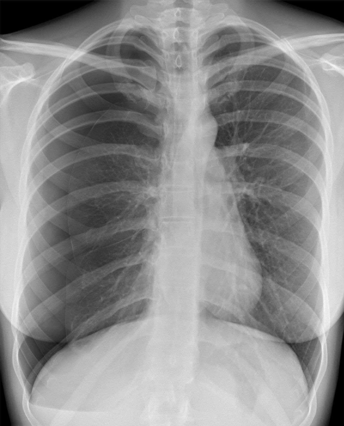
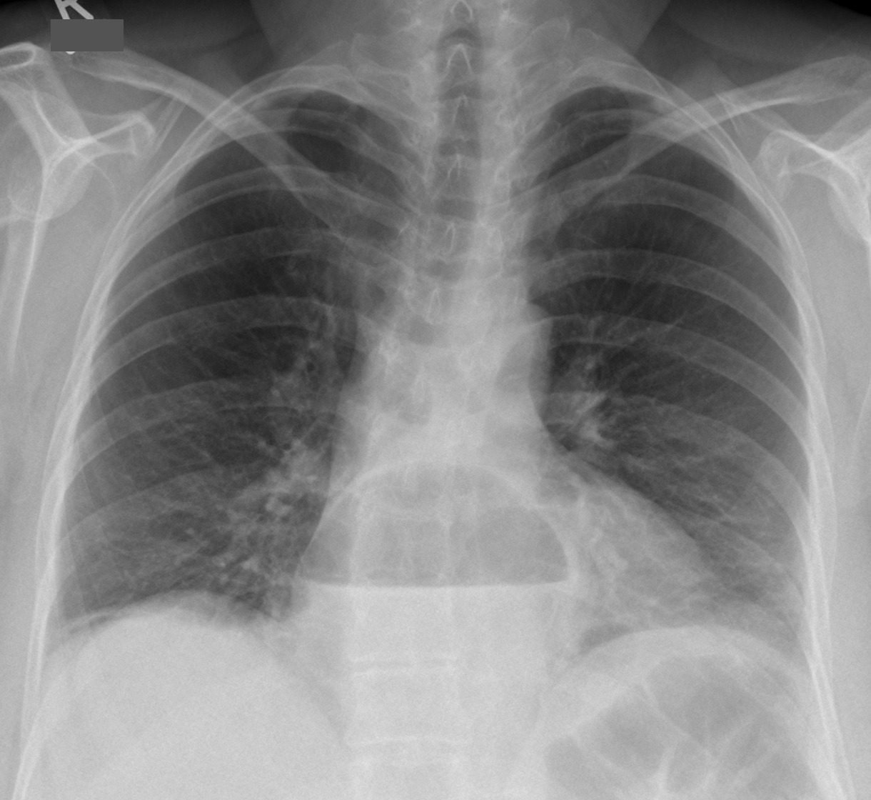
A 30-year-old woman fell off her bicycle 4 hours ago and has right-sided chest pain and mild dyspnea. HR 92 bpm, BP 124/78 mm Hg, SpO2 96% on room air. An upright PA chest radiograph is shown. Which of the following best describes the most important finding?
Hellerhoff, Wikimedia Commons, CC BY-SA 4.0 - ARight pulmonary contusion: ill-defined patchy opacity in the lateral right upper zone
- BRight rib fracture: cortical break of the right posterior 6th rib with a normal pleural space
- CRight pneumothorax: a thin visceral pleural line in the lateral right hemithorax with no lung markings peripheral to it
- DRight pleural effusion: blunted right costophrenic angle with a meniscus
- ENo acute abnormality: the curvilinear lines are breast and soft-tissue shadows
Show answer
Answer C
Source Felson's Principles of Chest Roentgenology, 5th ed. (2020); Fleischner Society Glossary of Terms for Thoracic Imaging (2024); image: Hellerhoff, Wikimedia Commons, CC BY-SA 4.0; author's caption (translated): right pneumothorax after a fall
- CX-002Multiple choiceRadiologyBasic
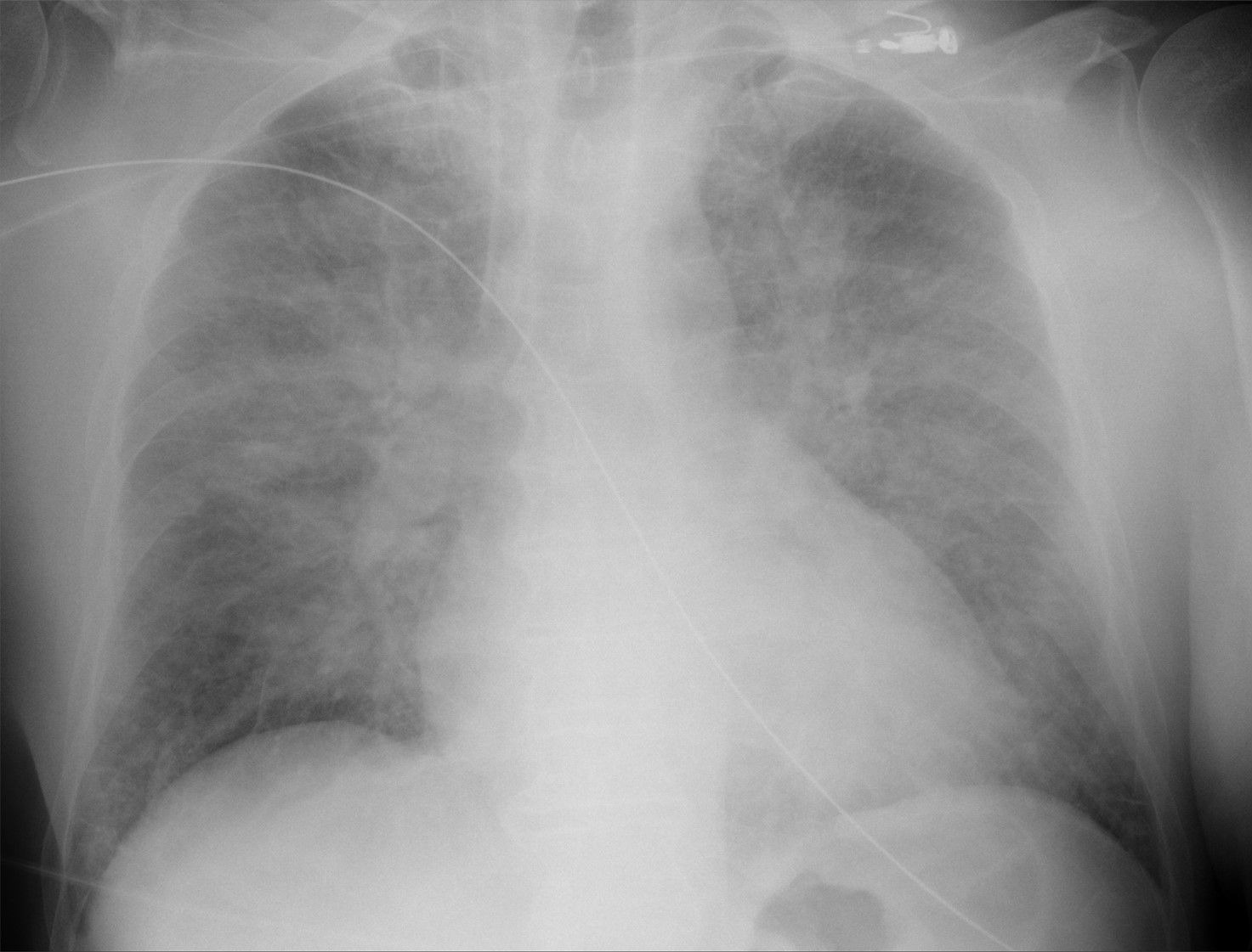
A 73-year-old man in the cardiac care unit on day 2 after an acute myocardial infarction develops sudden dyspnea; SpO2 is 85% on a face mask. A supine portable AP chest radiograph is shown. The best imaging diagnosis is:
Hellerhoff, Wikimedia Commons, CC BY-SA 4.0 - ABacterial pneumonia with lower-zone lobar consolidation
- BDiffuse miliary nodules of uniform size and density
- CPneumothorax with a visible visceral pleural line
- DBilateral large pleural effusions layering posteriorly on a supine film
- EBilateral perihilar-predominant airspace and interstitial opacities, consistent with cardiogenic pulmonary edema
Show answer
Answer E
Source Felson's Principles of Chest Roentgenology, 5th ed. (2020); image: Hellerhoff, Wikimedia Commons, CC BY-SA 4.0; author's caption (translated): pulmonary edema in cardiogenic shock
- CX-003Multiple choiceRadiologyIntermediate
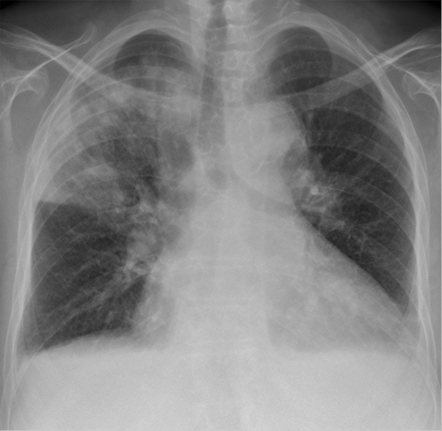
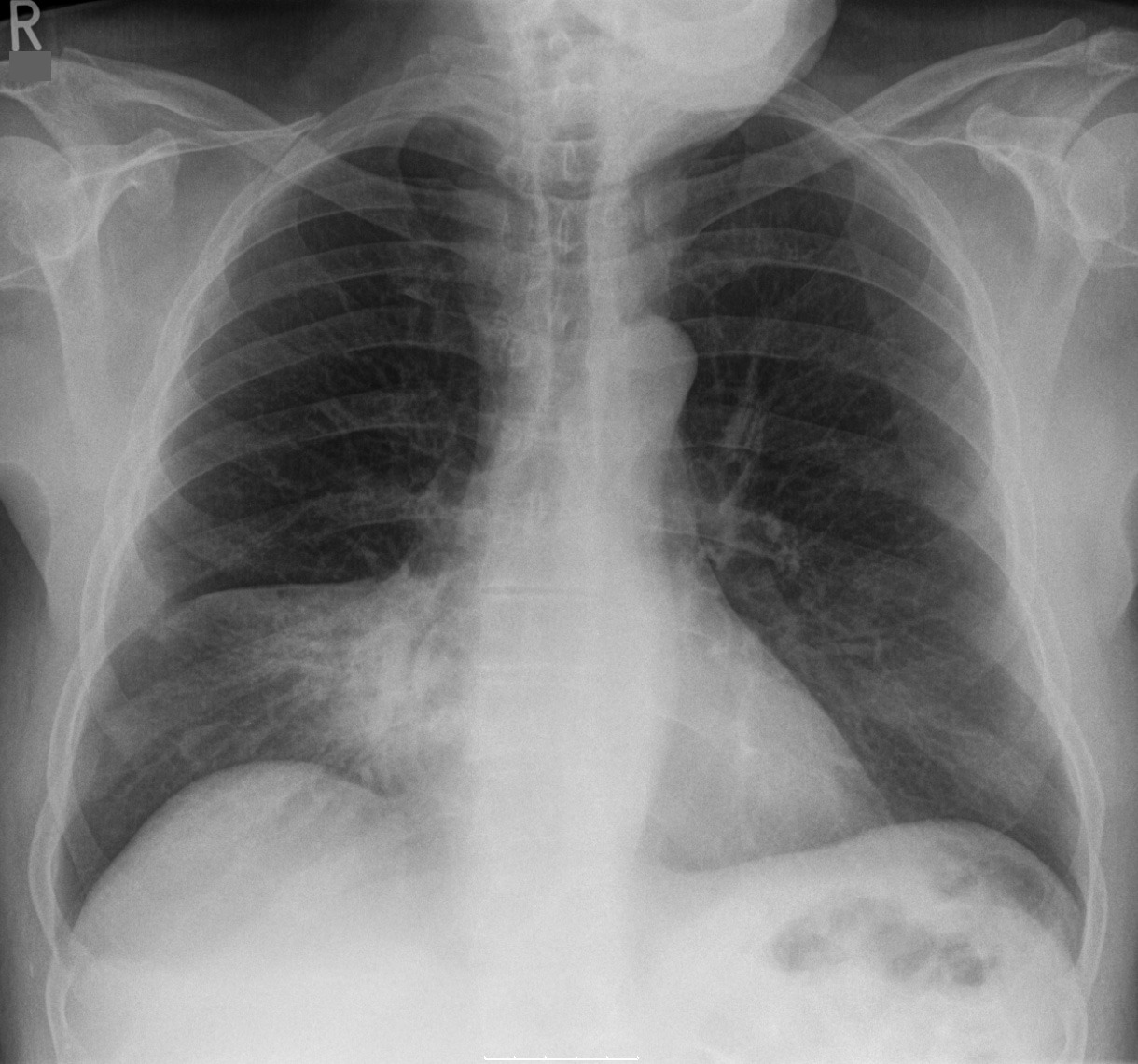
PA chest radiographs from two patients with community-acquired pneumonia are shown (image 1, image 2). The consolidation is most likely located in:
Hellerhoff, Wikimedia Commons, CC BY-SA 4.0 Hellerhoff, Wikimedia Commons, CC BY-SA 3.0 - AImage 1, right upper lobe; image 2, right middle lobe
- BImage 1, right upper lobe; image 2, right lower lobe
- CImage 1, right middle lobe; image 2, right upper lobe
- DImage 1, superior segment of the right lower lobe; image 2, right middle lobe
- EImage 1, right upper lobe; image 2, lingula
Show answer
Answer A
Source Felson's Principles of Chest Roentgenology, 5th ed. (2020) (silhouette sign); image: Hellerhoff, Wikimedia Commons, image 1 CC BY-SA 4.0; image 2 CC BY-SA 3.0; author's caption (translated): image 1, right upper lobe lobar pneumonia bounded inferiorly by the fissure; image 2, right middle lobe pneumonia
- CX-004Multiple choiceRadiologyAdvanced
A 66-year-old man on day 3 of mechanical ventilation in the ICU has a sudden fall in SpO2 from 95% to 86% with rising peak airway pressure. The tube depth at the teeth is unchanged from yesterday's radiograph, which showed the tip about 4 cm above the carina. A supine portable chest radiograph (standard orientation) is shown. The most likely diagnosis and best management are:
Hellerhoff, Wikimedia Commons, CC BY-SA 4.0 - ALeft lung atelectasis; intensify airway clearance, with bronchoscopic suctioning if needed
- BLeft tension pneumothorax; immediate needle decompression
- CExtensive left consolidation from progressive pneumonia; escalate antibiotics
- DLarge left hemothorax; chest tube and blood transfusion
- ELarge left pleural effusion; ultrasound-guided thoracentesis
Show answer
Answer A
Source Felson's Principles of Chest Roentgenology, 5th ed. (2020) (signs of volume loss); AARC Clinical Practice Guidelines: Artificial Airway Suctioning (Respir Care 2022;67:258–271); AARC CPG: Nonpharmacologic Airway Clearance Therapies in Hospitalized Patients (2013); image: Hellerhoff, Wikimedia Commons, CC BY-SA 4.0; author's caption (translated): subtotal left lung atelectasis from mucus plugging, with marked compensatory expansion of the right lung across the midline
- CX-005Short answerRadiologyIntermediate
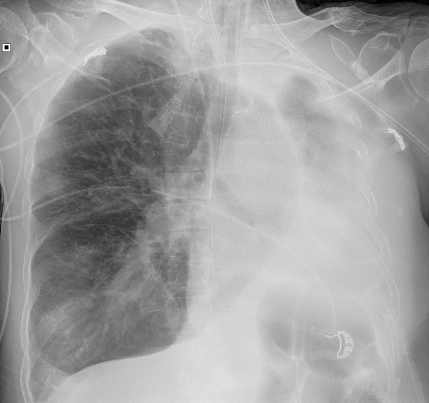
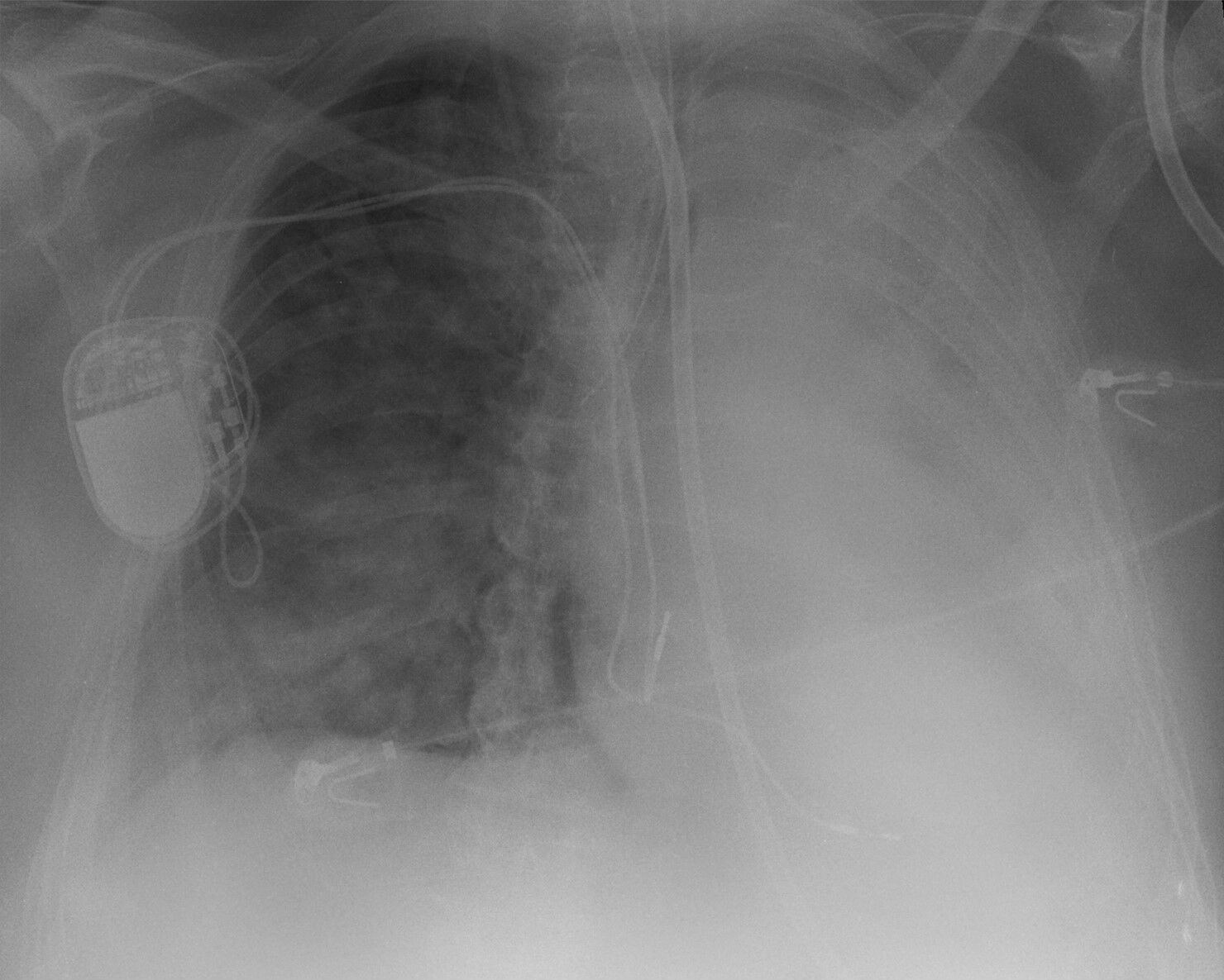
A 79-year-old man was intubated in the emergency department for respiratory failure and transferred to the ICU. SpO2 has gradually fallen from 95% after intubation to 87%, and peak airway pressure has risen. A portable chest radiograph is shown. Describe the main abnormalities and the management.
Hellerhoff, Wikimedia Commons, CC BY-SA 4.0 Rubric
- +3Identifies the endotracheal tube tip in the right main bronchus (tube too deep)
- +2Identifies left lung atelectasis, or explains that leftward mediastinal shift indicates volume loss
- +3Withdraws the tube so the tip is above the carina (about 3–7 cm, or T2–T4; the head-neutral qualifier is not required)
- +1Increases FiO2 and/or confirms with auscultation, end-tidal CO2, or a repeat radiograph
- +1Mentions the pacemaker, enteric tube, or any other device (side not required)
- -3Proposes a chest tube or thoracentesis as initial or primary management of the left opacity, or attributes it mainly to effusion, hemothorax, or pneumothorax (a contingent plan to look for effusion after repositioning is not penalized)
Show answer
Reference answer Findings: (1) the endotracheal tube is too deep, with its tip beyond the carina in the right main bronchus; (2) dense opacification of the left hemithorax from left lung atelectasis (the left lung is not ventilated), with the mediastinum and trachea shifted to the left (volume loss) and loss of the left heart border and hemidiaphragm; (3) also a pacemaker generator with transvenous leads (right chest wall, assuming standard orientation), an enteric tube, and ECG electrodes. Management: (1) increase FiO2 to 1.0 (or hand-ventilate with 100% oxygen) and withdraw the tube under auscultation so the tip is about 3–7 cm above the carina (ideally about 5 cm, roughly T2–T4, head neutral), resecure it, and document the depth at the teeth; (2) confirm symmetric breath sounds and end-tidal CO2 and repeat the radiograph to confirm position and left lung re-expansion; (3) suction and recruitment as needed; bronchoscopy if the left lung stays collapsed; if it still does not re-expand, ultrasound to look for a coexisting effusion is reasonable; (4) do not treat the left opacity as effusion or hemothorax with a chest tube first.
Source Felson's Principles of Chest Roentgenology, 5th ed. (2020); image: Hellerhoff, Wikimedia Commons, CC BY-SA 4.0; author's caption (translated): endotracheal tube malpositioned in the right main bronchus with left lung atelectasis
- CX-006Multiple choiceRadiologyBasic
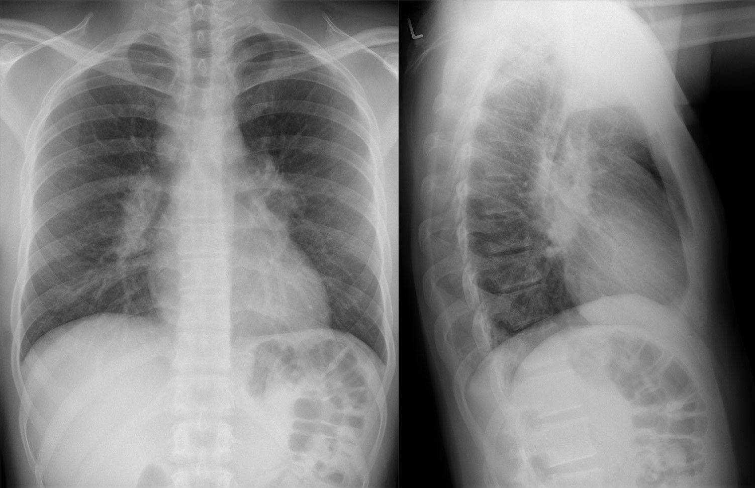
A 28-year-old asymptomatic man has PA and lateral chest radiographs for a pre-employment examination, shown here. Physical examination is normal. The most likely diagnosis is:
Hellerhoff, Wikimedia Commons, CC BY-SA 4.0 - APrimary tuberculosis
- BPulmonary arterial hypertension
- CSarcoidosis
- DLymphoma
- ECentral lung cancer
Show answer
Answer C
Source ATS Clinical Practice Guideline: Diagnosis and Detection of Sarcoidosis (2020); Felson's Principles of Chest Roentgenology, 5th ed. (2020); image: Hellerhoff, Wikimedia Commons, CC BY-SA 4.0; author's caption (translated): bilateral hilar lymphadenopathy in sarcoidosis
- CX-007Short answerRadiologyIntermediate
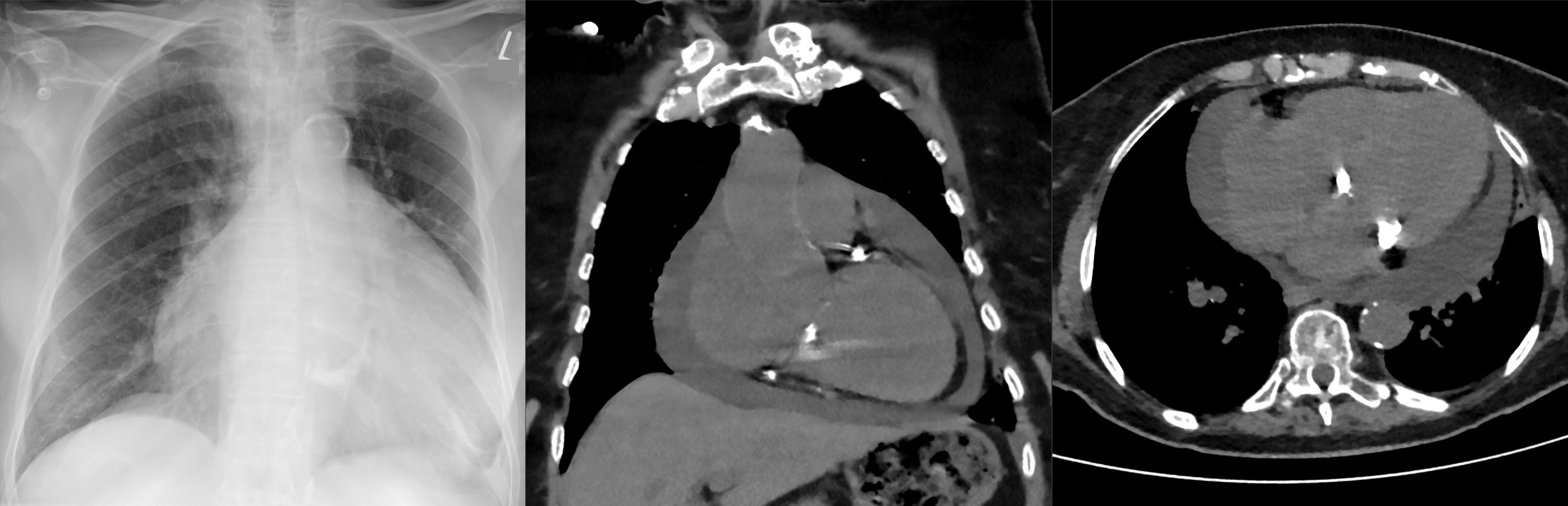
An 85-year-old woman has had progressive dyspnea for 3 weeks, worse lying flat; her past history is incomplete. BP 132/78 mm Hg, HR 96 bpm, no pulsus paradoxus, no obvious jugular venous distension, muffled heart sounds. Left: PA chest radiograph; middle and right: noncontrast chest CT, coronal and axial, from the same time. Give the imaging diagnosis with supporting findings, and the next tests and management.
Hellerhoff, Wikimedia Commons, CC BY-SA 4.0 Rubric
- +3Diagnoses pericardial effusion (2 points for effusion alone, 3 if described as moderate-to-large or large)
- +1Radiograph findings: flask-shaped silhouette without obvious pulmonary congestion
- +1CT findings: fluid-density band around the heart
- +2Chooses echocardiography first to size the effusion and assess for tamponade
- +2Gives at least two indications for pericardiocentesis (tamponade, symptomatic moderate-to-large effusion, suspected bacterial or neoplastic cause) and sends the fluid for analysis
- +1Lists at least two etiologic or baseline tests, or checks coagulation and excludes dissection before drainage
- -3Uses diuretics or vasodilators as the main treatment of the effusion (cautious diuresis for coexisting heart failure is not penalized)
Show answer
Reference answer Diagnosis: large pericardial effusion. Findings: on the radiograph the cardiac silhouette is markedly enlarged in both directions with a flask (water-bottle) shape and no obvious pulmonary venous congestion or interstitial edema, suggesting the enlargement is mostly fluid (the radiograph alone cannot distinguish this and cannot exclude coexisting cardiomegaly); CT shows a fluid-density band surrounding the heart, separated from the myocardium by the epicardial fat line. Next: (1) prompt echocardiography to size the effusion and look for tamponade (right atrial collapse in late diastole to early systole, right ventricular early diastolic collapse, plethoric IVC without respiratory variation, exaggerated respiratory variation of mitral and tricuspid inflow); (2) currently normal BP, no pulsus, no JVD — no classic tamponade, but monitor closely; (3) pericardiocentesis is indicated for tamponade, symptomatic moderate-to-large effusion not responding to medical therapy, or suspected bacterial or neoplastic cause; check coagulation and anticoagulant use first; noncontrast CT cannot exclude aortic dissection, and hemopericardium from dissection needs emergency surgery rather than pericardiocentesis; send fluid for cell count, chemistry, ADA, cytology, and culture; (4) etiologic and baseline tests: CRP/ESR, CBC, troponin, ECG, thyroid function, renal function, malignancy and tuberculosis workup, cardiac surgery or MI history, autoimmune disease. Diuretics and vasodilators are not the main treatment.
Source ESC Guidelines for the Diagnosis and Management of Pericardial Diseases (2015); ESC Guidelines for the Management of Myocarditis and Pericarditis (2025); image: Hellerhoff, Wikimedia Commons, CC BY-SA 4.0; author's caption (translated): pericardial effusion with the typical flask (Bocksbeutel) shape of the cardiac silhouette
- CX-008Short answerRadiologyIntermediate
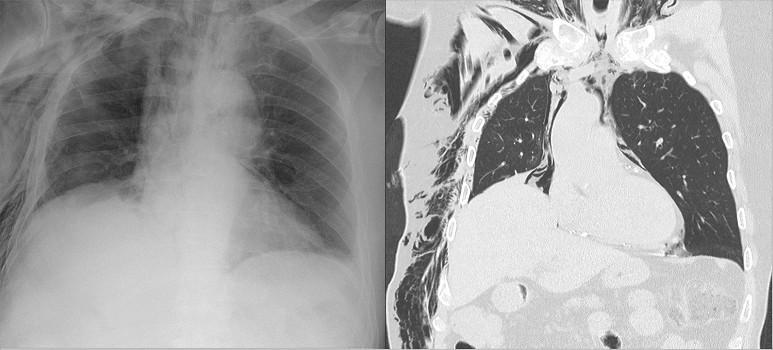
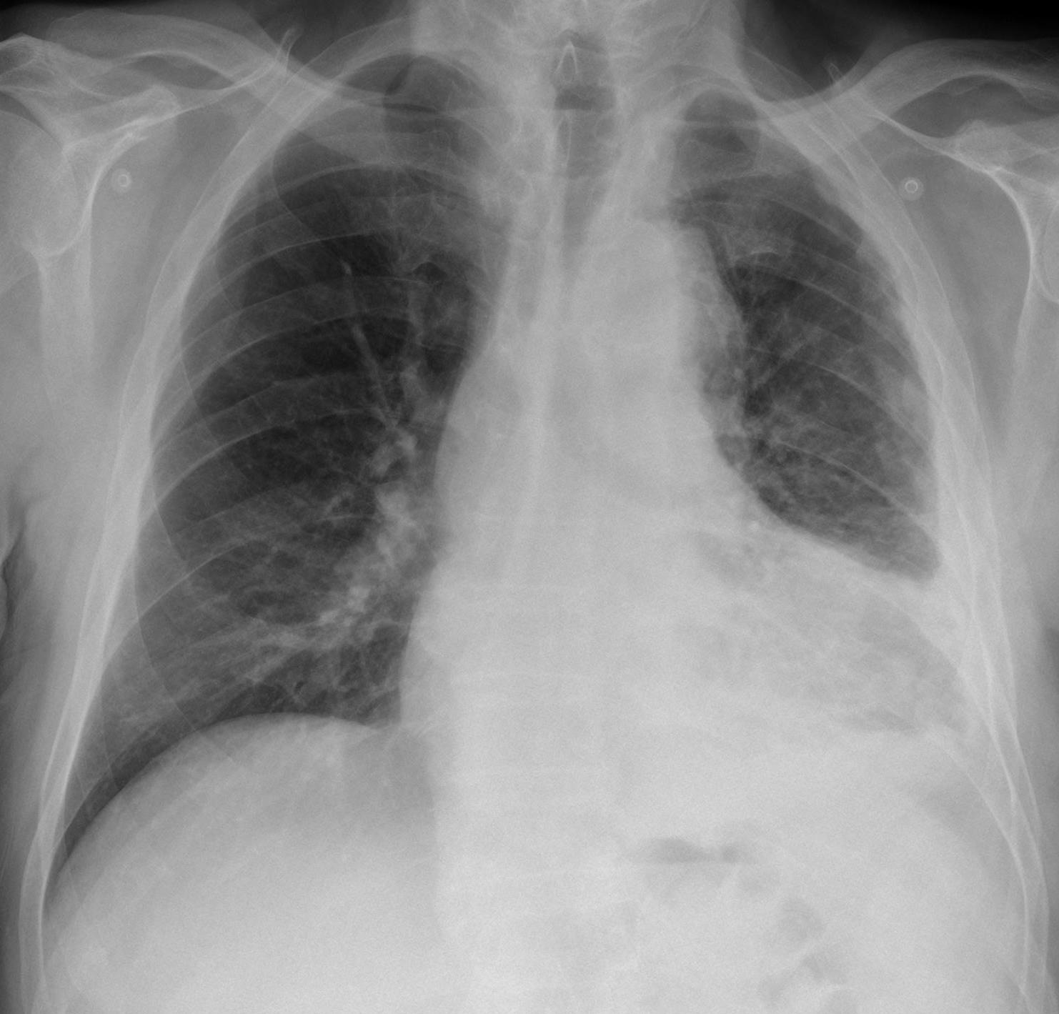
An 88-year-old man fell and sustained multiple right rib fractures. Left: portable chest radiograph; right: coronal chest CT reconstruction (lung window). Describe the abnormalities, the associated injuries to look for, and the key management points.
Hellerhoff, Wikimedia Commons, CC BY-SA 4.0 Rubric
- +2Identifies subcutaneous emphysema
- +2Identifies pneumomediastinum (CT findings suffice; discussing the distinction from pneumopericardium is acceptable)
- +2States that a pneumothorax is likely or must be excluded on the full CT (anterior, medial, subpulmonic); +1 if it asserts a pneumothorax is present without noting it is not clearly shown; 0 if it states there is no pneumothorax
- +1Raises concern for tracheobronchial or esophageal injury
- +1States chest tube indications (evident or enlarging pneumothorax, compromise) or the drain-versus-observation decision before positive-pressure ventilation
- +1Adequate analgesia (regional block or multimodal)
- +1Respiratory care (coughing, therapy, monitoring) with admission to a monitored bed for an older patient
- -2Calls the gas aortic dissection or pericardial effusion, or calls it pneumopericardium without considering pneumomediastinum
Show answer
Reference answer Findings: (1) extensive subcutaneous emphysema of the right chest wall and axilla extending into the neck; (2) pneumomediastinum: gas streaks around the mediastinal great vessels and trachea on CT, with a faint gas-outlined double line along the aortic arch and heart border on the radiograph (faint at this resolution; on CT, a thin gas line beneath the inferior heart surface is the correlate of the continuous diaphragm sign). Gas above the pericardial reflection, around the great vessels and trachea, is mediastinal rather than pericardial. Rib fractures are not well shown on this lung-window image and need bone windows or 3D reconstruction. Look for: pneumothorax — according to the source a pneumothorax was present, but it is not clearly shown on these two images; subcutaneous emphysema often indicates one, and an anterior, medial, or subpulmonic pneumothorax is easily missed on a supine portable film, so search the full CT; hemothorax; pulmonary contusion. Pneumomediastinum after blunt trauma usually results from alveolar rupture with gas tracking along the bronchovascular sheaths (Macklin effect); tracheobronchial or esophageal injury is uncommon but must not be missed (increasing gas, persistent air leak after chest tube, odynophagia: bronchoscopy or esophagography/CT). Management: analgesia (regional block or multimodal analgesia in older adults, accounting for anticoagulant or antiplatelet use) to allow coughing and deep breathing; chest tube for an evident or enlarging pneumothorax or respiratory or circulatory compromise; an occult pneumothorax seen only on CT may be observed even with positive-pressure ventilation if closely monitored (OPTICC), but drain it if it enlarges, causes compromise, or cannot be closely watched (e.g., long surgery or transfer); this much gas warrants a lower threshold for a drain; monitor SpO2 and work of breathing; respiratory therapy. Advanced age with multiple rib fractures carries high risk of pneumonia and respiratory failure; admit to a monitored bed.
Source Felson's Principles of Chest Roentgenology, 5th ed. (2020); EAST Practice Management Guideline: Pain Management in Blunt Thoracic Trauma (2016); OPTICC trial (Clements et al., Am J Surg 2021; occult pneumothorax); clinical scenario adapted; image: Hellerhoff, Wikimedia Commons, CC BY-SA 4.0; author's caption (translated): soft-tissue and mediastinal emphysema after multiple rib fractures; note the double line along the aortic arch and heart border; a pneumothorax was also present
- CX-009Multiple choiceRadiologyBasic
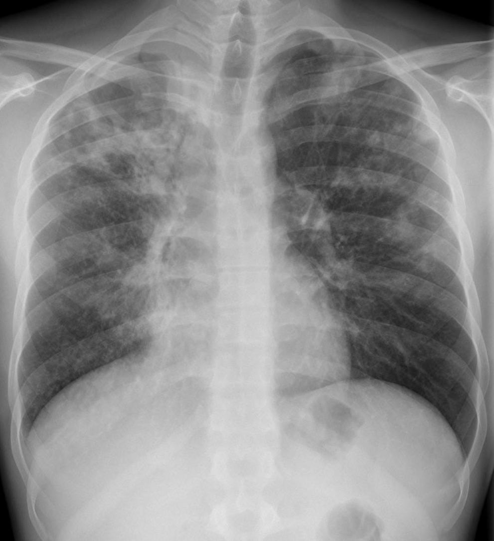
A 27-year-old man has had a productive cough for 2 months; his HIV status is unknown. A PA chest radiograph is shown. The best assessment and first step are:
Hellerhoff, Wikimedia Commons, CC BY-SA 4.0 - ARight upper lobe lung abscess: IV antibiotics covering anaerobes and a repeat radiograph in 2 weeks
- BCentral right upper lobe lung cancer with obstructive pneumonia: bronchoscopic biopsy first
- CActive pulmonary tuberculosis: start empiric RIPE therapy immediately without waiting for microbiology
- DActive pulmonary tuberculosis: airborne isolation, sputum rapid molecular testing (e.g., Xpert MTB/RIF Ultra) plus AFB smear and culture
- EActive pulmonary tuberculosis: CT-guided percutaneous lung biopsy first to confirm the diagnosis
Show answer
Answer D
Source WHO Consolidated Guidelines on Tuberculosis, Module 3: Diagnosis (2024); ATS/IDSA/CDC Clinical Practice Guidelines: Diagnosis of Tuberculosis in Adults and Children (2017); CDC Guidelines for Preventing the Transmission of Mycobacterium tuberculosis in Health-Care Settings (2005); image: Hellerhoff, Wikimedia Commons, CC BY-SA 4.0; author's caption (translated): active cavitary tuberculosis
- CX-010Multiple choiceRadiologyAdvanced
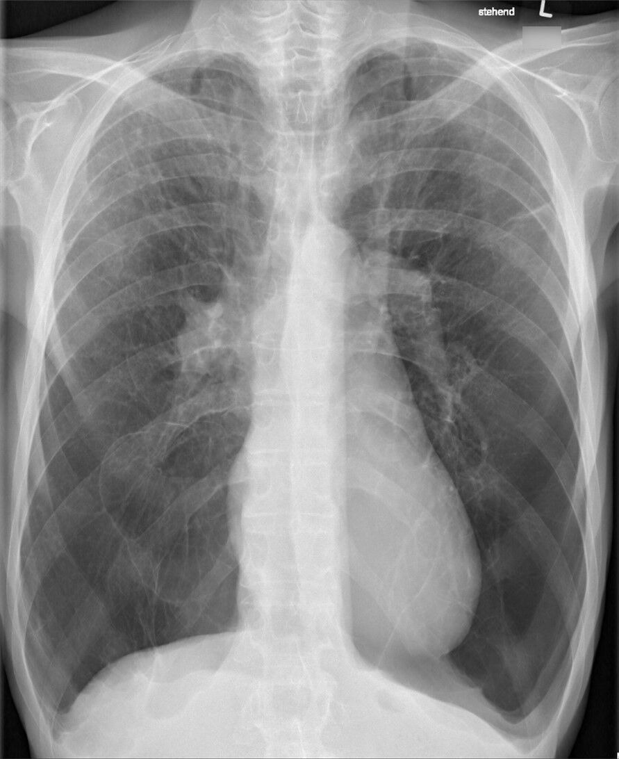
A 62-year-old man has had progressive exertional dyspnea for 4 years. An upright PA chest radiograph is shown. Regarding the distribution of emphysema and its most likely cause:
Hellerhoff, Wikimedia Commons, CC BY-SA 4.0 - AUpper-zone predominant emphysema, smoking-related centrilobular type
- BUnilateral hyperlucency with reduced vessels, Swyer-James syndrome
- CGiant bilateral upper-lobe bullae, bullous lung disease
- DLower-zone predominant emphysema, alpha-1 antitrypsin deficiency
- ERight pneumothorax with lung collapse
Show answer
Answer D
Source GOLD 2025 Report; ATS/ERS Statement: Standards for the Diagnosis and Management of Individuals With Alpha-1 Antitrypsin Deficiency (2003); image: Hellerhoff, Wikimedia Commons, CC BY-SA 4.0; author's caption (translated): severe emphysema in alpha-1 antitrypsin deficiency
- CX-011Multiple choiceRadiologyAdvanced
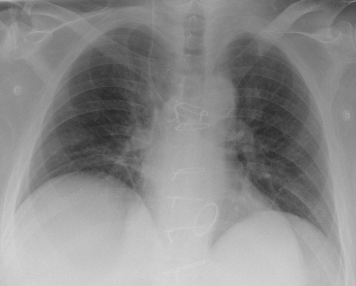
A 50-year-old man after cardiac surgery has a central venous catheter placed through the right internal jugular vein; a routine portable radiograph after placement is shown. Regarding the catheter tip position, the best assessment is:
Hellerhoff, Wikimedia Commons, CC BY-SA 4.0 - AAt the cavoatrial junction; ready to use
- BTurns medially at the level just above the right main bronchus, in the azygos vein; reposition
- CIn the right ventricle; withdraw
- DCrossed the midline into the left brachiocephalic vein; reposition
- EFollowed the common carotid artery into the aortic arch; remove and apply pressure
Show answer
Answer B
Source Felson's Principles of Chest Roentgenology, 5th ed. (2020) (line position); image: Hellerhoff, Wikimedia Commons, CC BY-SA 4.0; author's caption (translated): jugular central venous catheter malpositioned in the azygos vein; a left brachiocephalic position would project higher and rise toward the left
- CX-012Short answerRadiologyAdvanced
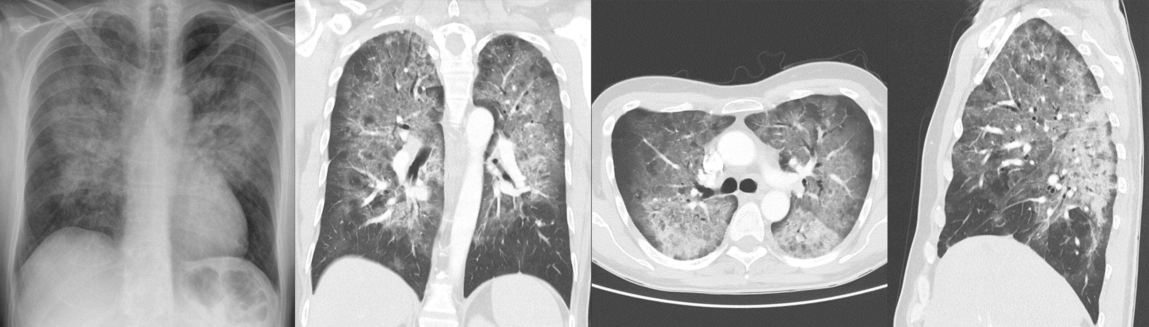
A 53-year-old man has had dry cough and exertional dyspnea for 3 weeks, low-grade fever, and 7 kg (15 lb) weight loss over 2 months. He has white oral plaques, a lymphocyte count of 600/μL, and LDH 452 U/L. SpO2 is 92% at rest and falls to 83% with exertion. Shown are a chest radiograph and same-day chest CT (coronal, axial, and sagittal, lung window). Give the main imaging findings, the most likely diagnosis, the immediate workup, and initial treatment.
Hellerhoff, Wikimedia Commons, CC BY-SA 4.0 Rubric
- +1Describes diffuse bilateral ground-glass opacity
- +1Adds any of: central or upper-mid lung predominance, geographic pattern, septal thickening
- +2Diagnoses Pneumocystis jirovecii pneumonia
- +1Orders HIV testing (with CD4 count)
- +1Induced sputum or BAL for P. jirovecii
- +2Chooses trimethoprim-sulfamethoxazole with dose or 21-day duration
- +2Adds corticosteroids within 72 hours when PaO2 <70 mm Hg or A-a gradient ≥35 mm Hg
- -3Treats as bacterial pneumonia with a beta-lactam alone, or uses corticosteroids alone
Show answer
Reference answer Imaging: the radiograph shows diffuse, roughly symmetric ground-glass and reticular opacities, perihilar and mid-upper zone predominant, with clear costophrenic angles. CT shows diffuse ground-glass opacity, central and upper-mid lung predominant with relative basal sparing, in a geographic pattern (some secondary lobules spared), possibly with interlobular septal thickening and small consolidations; no cavities, nodules, pneumothorax, or effusion. With subacute course, dry cough, exertional hypoxemia, thrush, lymphopenia, and elevated LDH, the most likely diagnosis is Pneumocystis jirovecii pneumonia (PCP); HIV infection must be considered first. Differential: CMV or other viral pneumonia (including COVID-19), hypersensitivity pneumonitis, pulmonary alveolar proteinosis, drug-induced lung injury. Workup: HIV testing (with CD4 count and viral load); induced sputum or BAL for P. jirovecii (stain, immunofluorescence, or PCR); arterial blood gas (PaO2, A-a gradient); serum (1,3)-beta-D-glucan. Treatment: trimethoprim-sulfamethoxazole (trimethoprim 15–20 mg/kg/day in 3–4 divided doses) for 21 days; if PaO2 <70 mm Hg on room air or A-a gradient ≥35 mm Hg, add prednisone within 72 hours of starting PCP therapy (40 mg twice daily for 5 days, 40 mg daily for 5 days, then 20 mg daily for 11 days); with a resting SpO2 of 92% he will likely qualify once the blood gas confirms it. If HIV is confirmed, start antiretroviral therapy within 2 weeks of starting PCP treatment, followed by secondary prophylaxis.
Source NIH/CDC/HIVMA-IDSA Guidelines for the Prevention and Treatment of Opportunistic Infections in Adults and Adolescents With HIV: Pneumocystis Pneumonia (current online version, accessed November 2026); image: Hellerhoff, Wikimedia Commons, CC BY-SA 4.0; author's caption (translated): Pneumocystis jirovecii pneumonia in a patient with HIV infection
- CX-013Multiple choiceRadiologyBasic
A 58-year-old woman has a screening PA chest radiograph, shown here. The retrocardiac abnormality is most likely:
Hellerhoff, Wikimedia Commons, CC BY-SA 3.0 - ALung abscess: left lower lobe cavity with an air-fluid level
- BHiatal hernia: stomach herniated into the posterior mediastinum through the esophageal hiatus, with an air-fluid level
- CPericardial cyst: smooth cystic opacity at the right cardiophrenic angle
- DPosterior mediastinal neurogenic tumor: well-defined paraspinal soft-tissue mass
- ELoculated pleural effusion: fusiform opacity within a fissure
Show answer
Answer B
Source Felson's Principles of Chest Roentgenology, 5th ed. (2020); image: Hellerhoff, Wikimedia Commons, CC BY-SA 3.0; author's caption (translated): large hiatal hernia with an air-fluid level on a chest radiograph
- CX-014Multiple choiceRadiologyAdvanced
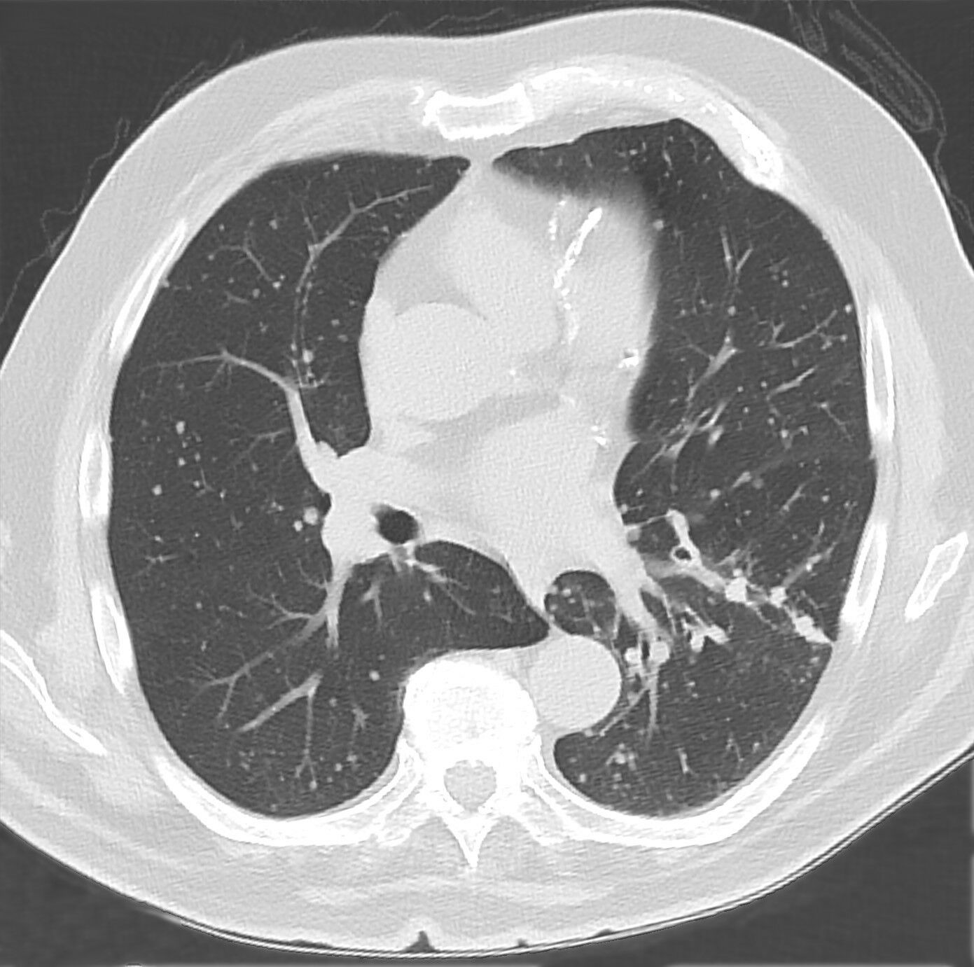
A 76-year-old man has had fatigue and weight loss for 3 months without fever. Chest CT (lung window) is shown. The best assessment and next step are:
Hellerhoff, Wikimedia Commons, CC BY-SA 4.0 - AMultiple randomly distributed nodules of varying size, hematogenous metastases; search for a primary tumor
- BPerilymphatic nodules along bronchovascular bundles and fissures, sarcoidosis; bronchoscopic biopsy
- CCentrilobular tree-in-bud opacities, infectious bronchiolitis; treat and repeat imaging
- DDiffuse uniform miliary nodules, miliary tuberculosis; start four-drug therapy immediately
- ECentrilobular ground-glass nodules, hypersensitivity pneumonitis; take a detailed exposure history
Show answer
Answer A
Source Fleischner Society Glossary of Terms for Thoracic Imaging (2024); Felson's Principles of Chest Roentgenology, 5th ed. (2020); image: Hellerhoff, Wikimedia Commons, CC BY-SA 4.0; author's caption (translated): multiple lung metastases from papillary thyroid carcinoma
- CX-015Short answerRadiologyAdvanced
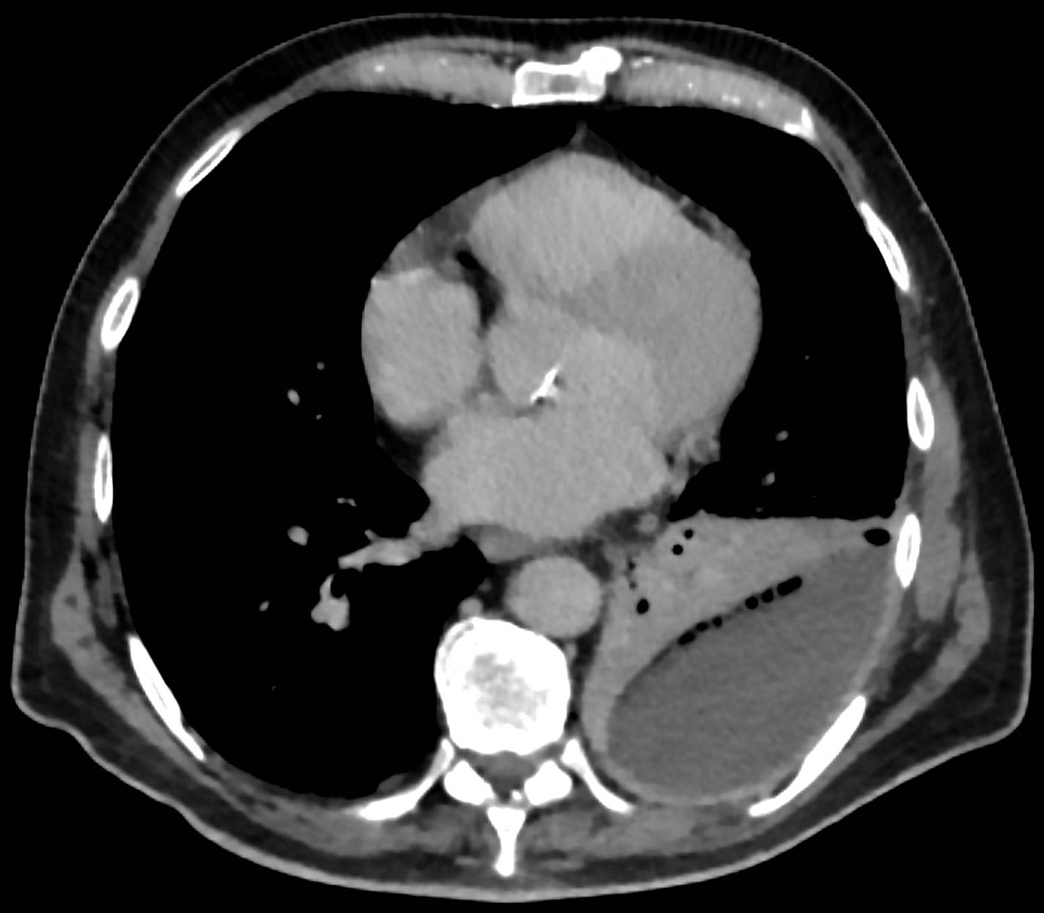
A 66-year-old man has had fever and left chest pain for 10 days; oral antibiotics for community-diagnosed pneumonia have failed. He has had no thoracentesis or surgery. WBC 18,000/μL, CRP 210 mg/L (21 mg/dL). An axial contrast-enhanced chest CT (venous phase, mediastinal window) is shown. Describe the imaging findings, the diagnosis, and the management plan.
Hellerhoff, Wikimedia Commons, CC BY-SA 4.0 Rubric
- +1Describes a left loculated (lenticular) collection
- +1Describes gas or bubbles within the pleural collection (air bronchograms in the consolidated lung alone earn no credit)
- +2Diagnoses pleural infection, empyema, or complicated parapneumonic effusion
- +2Drainage: chest tube (immediately, or after diagnostic aspiration meeting criteria) or primary surgical (VATS) drainage
- +1Sends pleural fluid for any of pH, glucose, Gram stain, or culture (credit also for draining frank pus without measuring pH)
- +1Antibiotics covering anaerobes (adjusted to culture)
- +1Intrapleural tPA and DNase together for residual fluid with uncontrolled infection, or surgical evaluation for VATS (no credit for streptokinase, tPA alone, DNase alone, or generic fibrinolytics without DNase)
- -3Only adjusts antibiotics (oral or IV) without proposing any drainage (not applied to a primary VATS plan)
Show answer
Reference answer Imaging: posterior left biconvex, lenticular loculated fluid collection with a broad base on the chest wall that compresses the adjacent lung (favoring a pleural process over lung abscess); a string of small gas bubbles along its anterior margin (pleural gas; gas within the consolidated lung on the mediastinal side is an air bronchogram, not to be confused); at most mild pleural enhancement at the margin; adjacent left lower lobe consolidation. Diagnosis: pleural infection — presumed empyema or complicated parapneumonic effusion, to be confirmed by aspiration — with left lower lobe pneumonia. Gas without prior instrumentation suggests gas-forming (anaerobic) infection or bronchopleural fistula; on the left, also exclude esophageal rupture. Drainage is expected here: a large loculated collection with gas in a septic patient. Management: (1) ultrasound-guided aspiration — if frank pus is aspirated, place a chest tube without measuring pH; otherwise send pH, glucose, LDH, Gram stain and culture (plus blood cultures). Drain if pH <7.2 (glucose <60 mg/dL [3.3 mmol/L] when pH is unavailable) or Gram stain or culture is positive; pH 7.2–7.4 is a gray zone, where LDH >900 IU/L favors drainage. (2) Chest tube (small bore usually suffices) with regular flushes. (3) IV antibiotics covering community pathogens and anaerobes (e.g., ampicillin-sulbactam, or a cephalosporin plus metronidazole), avoiding aminoglycosides, adjusted to cultures, switching to oral when improving; typically at least 2–3 weeks in total, guided by clinical, radiologic, and CRP response. (4) If fluid persists with uncontrolled infection after drainage, intrapleural tPA 10 mg plus DNase 5 mg twice daily for 3 days (MIST2 regimen), or thoracic surgery evaluation for VATS debridement or decortication. (5) Nutritional support.
Source BTS Guideline for Pleural Disease (2023); MIST2 (Rahman NM et al., N Engl J Med 2011); image: Hellerhoff, Wikimedia Commons, CC BY-SA 4.0; author's caption (translated): pleural empyema with adjacent consolidated lung and gas inclusions
- CX-016Multiple choiceRadiologyIntermediate
An 85-year-old retired shipyard worker who lives independently (ECOG performance status 1) has had persistent left chest pain for 3 months and weight loss, and wishes to pursue diagnosis and treatment. A PA chest radiograph is shown. Thoracentesis yields a bloody exudate; two cytology samples are negative. The most appropriate next step is:
Hellerhoff, Wikimedia Commons, CC BY-SA 4.0 - ATwo more cytology samples; if still negative, malignancy is excluded
- BContrast-enhanced CT, then image-guided or thoracoscopic pleural biopsy
- CEmpiric antituberculous therapy for 4 weeks and observe the effusion
- DTube drainage and pleurodesis without a tissue diagnosis
- EPET-CT; a positive scan establishes malignancy without biopsy
Show answer
Answer B
Source BTS Guideline for the Investigation and Management of Malignant Pleural Mesothelioma (2018); BTS Guideline for Pleural Disease (2023); image: Hellerhoff, Wikimedia Commons, CC BY-SA 4.0; author's caption (translated): pleural mesothelioma on a chest radiograph: circumferential left pleural thickening with effusion
- CX-017Multiple choiceRadiologyBasic
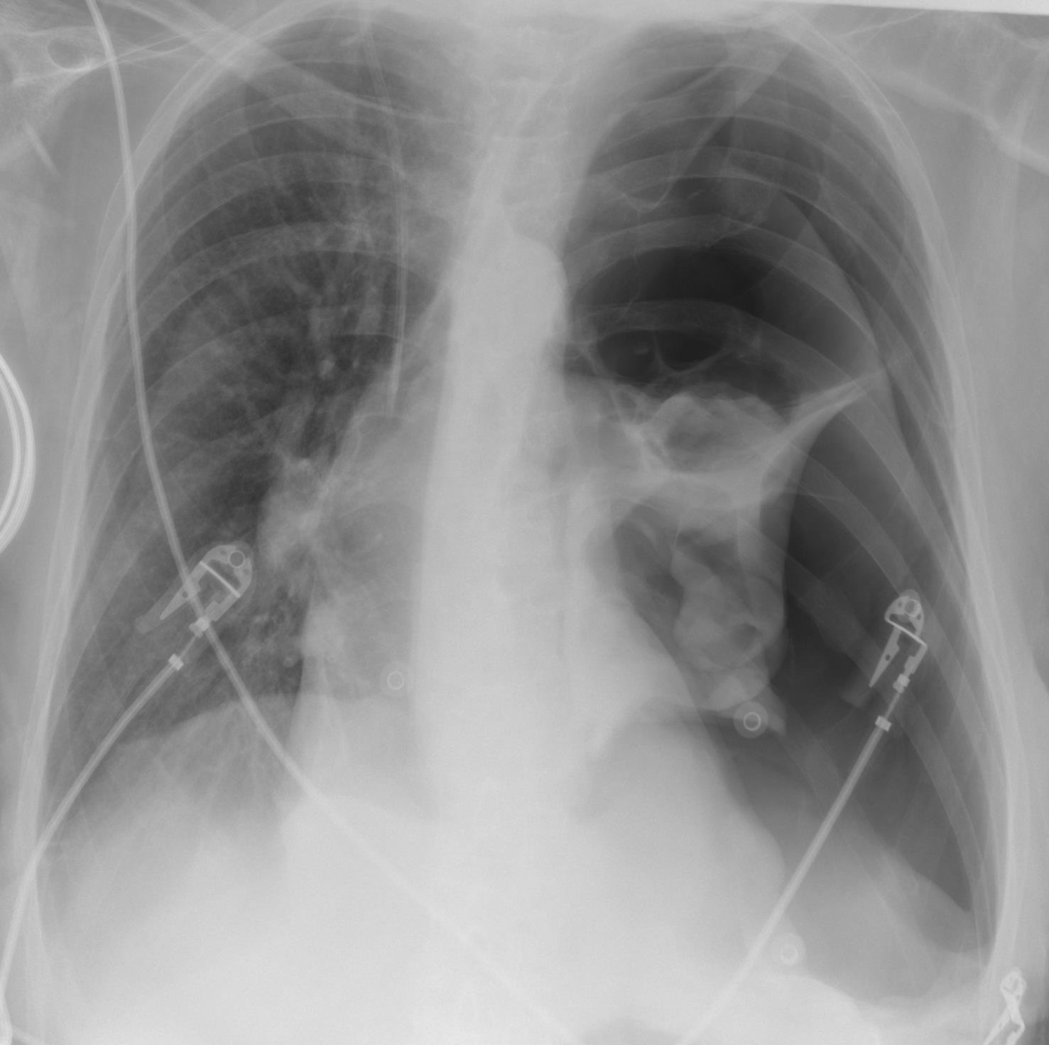
A 76-year-old woman in the ICU, breathing spontaneously, had an unsuccessful attempt at left subclavian venous catheterization, after which a right internal jugular catheter was placed. She then develops sudden dyspnea; BP falls to 72/40 mm Hg, HR 128 bpm, SpO2 82%. A chest radiograph obtained moments before the deterioration is shown. The most appropriate management is:
Hellerhoff, Wikimedia Commons, CC BY-SA 4.0 - ABronchoscopic suctioning of a left main bronchus mucus plug causing atelectasis
- BImmediate right-sided decompression (needle at the 4th/5th intercostal space anterior to the midaxillary line, or finger thoracostomy), followed by a right chest tube
- CIncrease supplemental oxygen (high-flow nasal cannula) and repeat the radiograph in 30 minutes
- DRapid fluids and norepinephrine, with CT once BP is stable
- EImmediate left-sided decompression (needle at the 4th/5th intercostal space anterior to the midaxillary line, or finger thoracostomy), followed by a left chest tube
Show answer
Answer E
Source BTS Guideline for Pleural Disease (2023); ATLS Advanced Trauma Life Support, 10th ed. (2018); clinical scenario adapted; image: Hellerhoff, Wikimedia Commons, CC BY-SA 4.0; author's caption (translated): left tension pneumothorax
- CX-018Multiple choiceRadiologyIntermediate
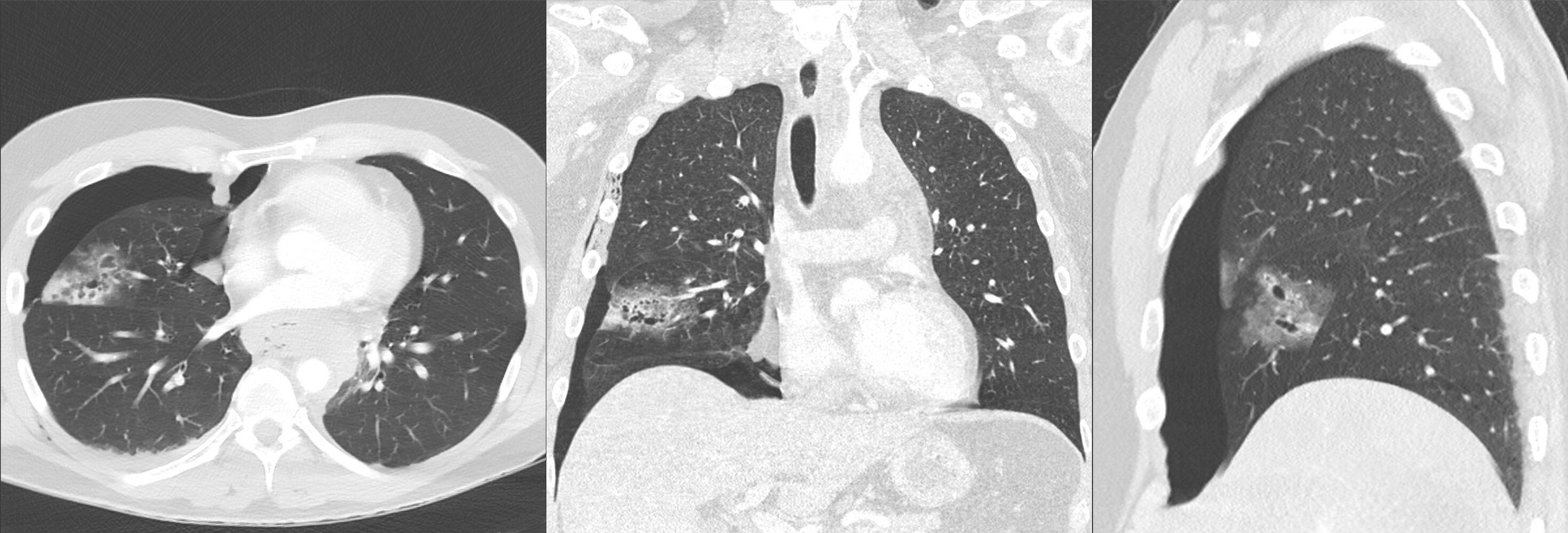
A 38-year-old man was in a car crash. Chest CT obtained on arrival in the emergency department, before chest tube placement, is shown (axial, coronal, sagittal; lung window); the right pneumothorax was then drained and the lung re-expanded. On day 2 he has right chest pain and small-volume blood-streaked sputum; vital signs are stable and he is afebrile. Regarding the right middle lobe lesion, the most likely diagnosis and management are:
Hellerhoff, Wikimedia Commons, CC BY-SA 4.0 - ALung abscess: IV antibiotics, percutaneous drainage if needed
- BCavitating lung cancer: CT-guided percutaneous biopsy
- CContusion with traumatic pneumatoceles: supportive care, analgesia, pulmonary toilet
- DInfected pulmonary sequestration: elective surgical resection
- ECavitary tuberculosis: sputum testing, antituberculous therapy
Show answer
Answer C
Source Felson's Principles of Chest Roentgenology, 5th ed. (2020) (course of traumatic pneumatoceles); EAST Practice Management Guideline: Pulmonary Contusion and Flail Chest (Simon B et al., J Trauma Acute Care Surg 2012;73:S351–S361); clinical scenario adapted; image: Hellerhoff, Wikimedia Commons, CC BY-SA 4.0; author's caption (translated): pulmonary contusion and laceration, mainly of the middle lobe, with pneumothorax and traumatic pneumatoceles
- CX-019Short answerRadiologyAdvanced
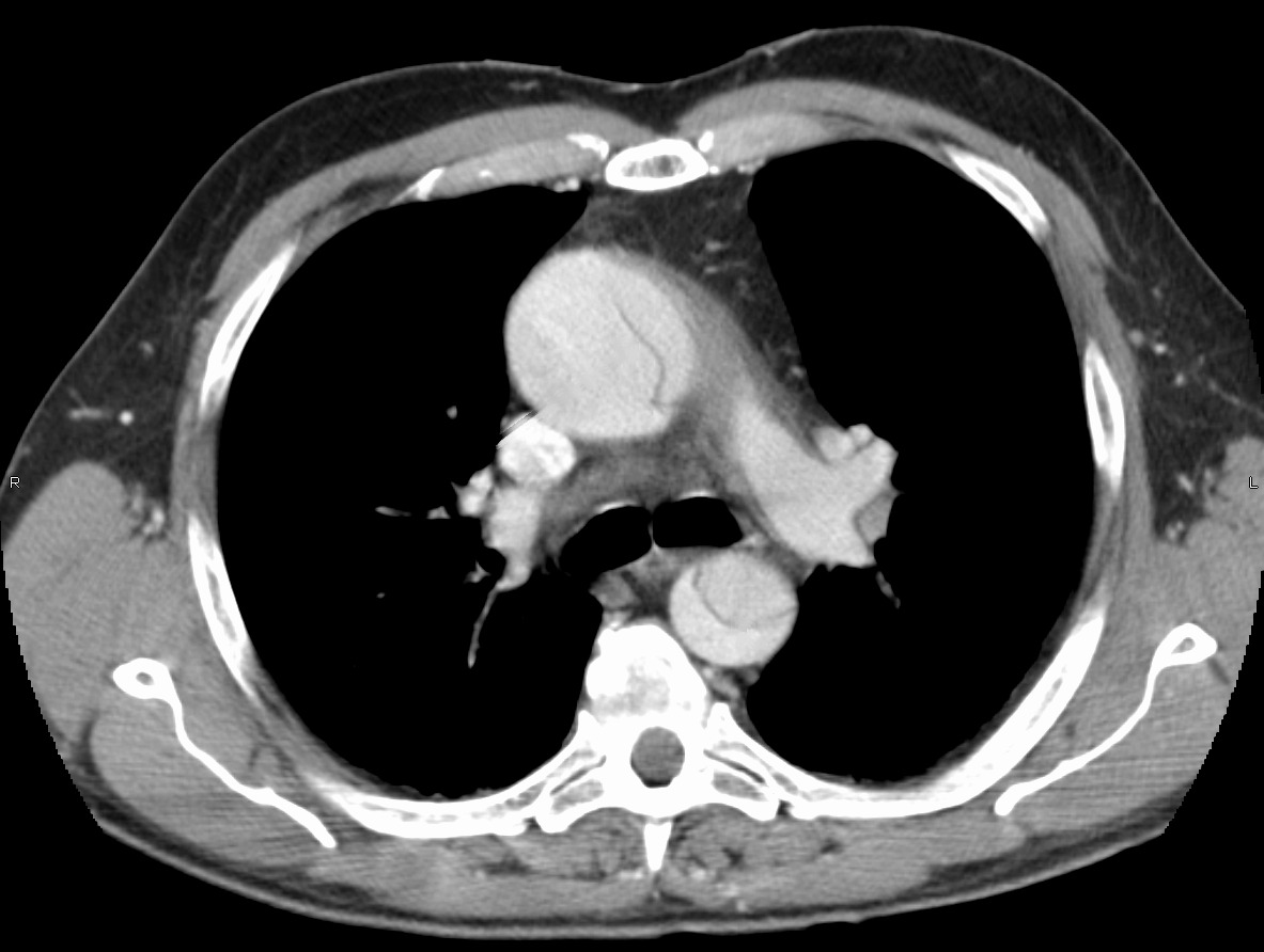
A 62-year-old man has sudden tearing chest and back pain for 2 hours. BP 178/96 mm Hg in the right arm and 150/88 mm Hg in the left, HR 102 bpm. A contrast-enhanced axial chest CT is shown. Describe the imaging findings, classification, emergency management before definitive treatment, and what must be avoided.
James Heilman, MD, Wikimedia Commons, CC BY-SA 3.0; arrows removed by BenchClub Rubric
- +1Identifies an intimal flap in the ascending aorta
- +1Also identifies a flap or dual lumen in the descending aorta
- +2Classifies as Stanford type A (recognizing ascending involvement suffices)
- +2Emergency cardiac surgery, or emergency transfer to an aortic or cardiac surgical center
- +2Anti-impulse therapy: +1 beta-blocker first targeting HR ≤60 bpm; +1 then systolic BP about 100–120 mm Hg (accept <120)
- +1IV opioid analgesia, or assessment for at least one complication (tamponade, aortic regurgitation, coronary involvement, malperfusion)
- +2States what to avoid: anticoagulant, thrombolytic, or antiplatelet therapy, or a vasodilator before rate control (either earns credit)
- -3(applied once) Gives anticoagulant, thrombolytic, or antiplatelet therapy, or lowers BP with a vasodilator alone before rate control
Show answer
Reference answer Imaging: at this level, linear low-attenuation intimal flaps in both the ascending and descending aorta divide each lumen into true and false lumens. Classification: involvement of the ascending aorta makes this Stanford type A; with descending involvement it is consistent with DeBakey type I (the full extent requires the complete CTA). Management: (1) immediate cardiac surgery consultation, or emergency transfer to an aortic surgical center; emergency surgery is definitive. (2) Anti-impulse therapy: IV beta-blocker (esmolol or labetalol) first to bring HR to about 60 bpm or less, then a vasodilator (e.g., nicardipine or nitroprusside) as needed to bring systolic BP to about 100–120 mm Hg while maintaining organ perfusion, titrating to the higher-reading (right) arm; a nondihydropyridine calcium channel blocker if beta-blockers are contraindicated. (3) IV opioid analgesia. (4) Assess complications: pericardial effusion and tamponade, aortic regurgitation (echocardiography), coronary involvement (ECG), cerebral, renal, mesenteric, or limb malperfusion. Avoid: anticoagulants, thrombolytics, and antiplatelet agents (do not treat as an acute coronary syndrome); vasodilator alone without prior rate control (reflex tachycardia propagates the dissection); blind large-volume pericardiocentesis for tamponade.
Source ACC/AHA Guideline for the Diagnosis and Management of Aortic Disease (2022); ESC Guidelines for the Management of Peripheral Arterial and Aortic Diseases (2024); image: James Heilman, MD, Wikimedia Commons, CC BY-SA 3.0; author's caption (translated): type A aortic dissection (modified: annotation arrows removed)