@joshua/dermatology-images
Dermatology: reading skin lesions
Author-made · not reviewedDermatologyCC BY 4.0
Written by the BenchClub team with AI help. Answers follow public guidelines and textbooks; not yet reviewed by a practising physician.
v119 questions19 with imagesUpdated Nov 17, 2026
- DR-001Multiple choiceDermatologyBasic
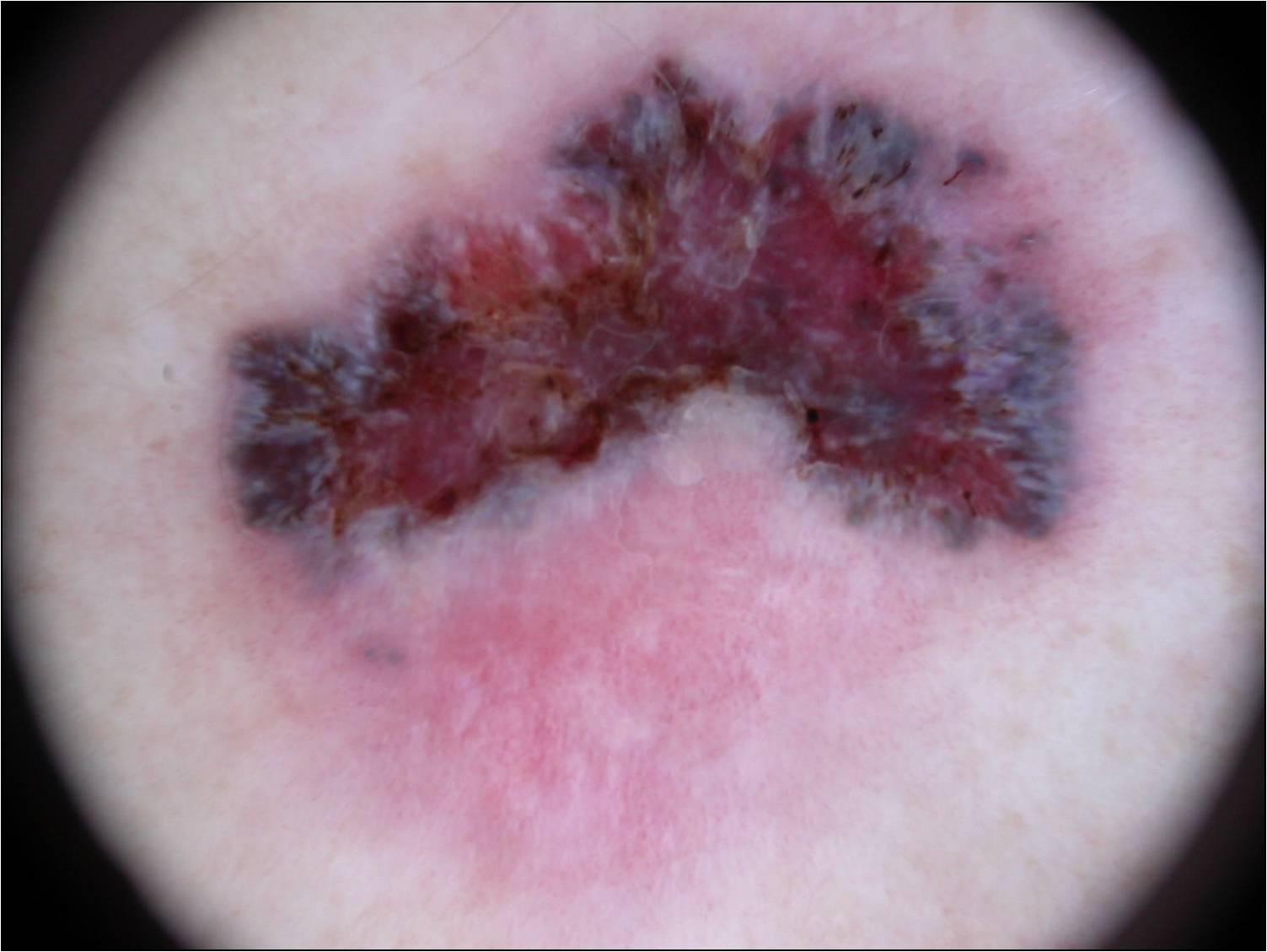
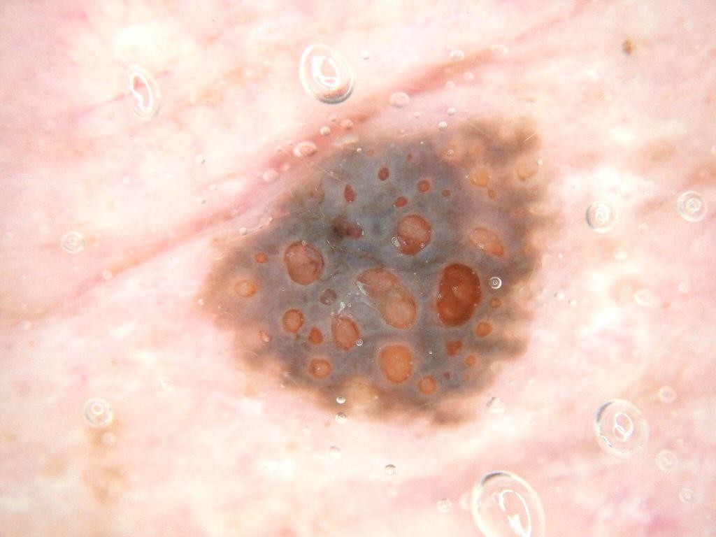
A 55-year-old woman points out a lesion on her lower leg that has changed in color over the past year. The dermoscopic image is shown. What is the most likely diagnosis?
Anonymous contributor, ISIC Archive ISIC_0000030, CC0 - ASeborrheic keratosis
- BPigmented basal cell carcinoma
- CMelanoma
- DHemangioma
- EDermatofibroma
Show answer
Answer C
Source Kittler H et al. Standardization of terminology in dermoscopy/dermatoscopy, JAAD 2016; ISIC Archive histopathology label
- DR-002Multiple choiceDermatologyIntermediate
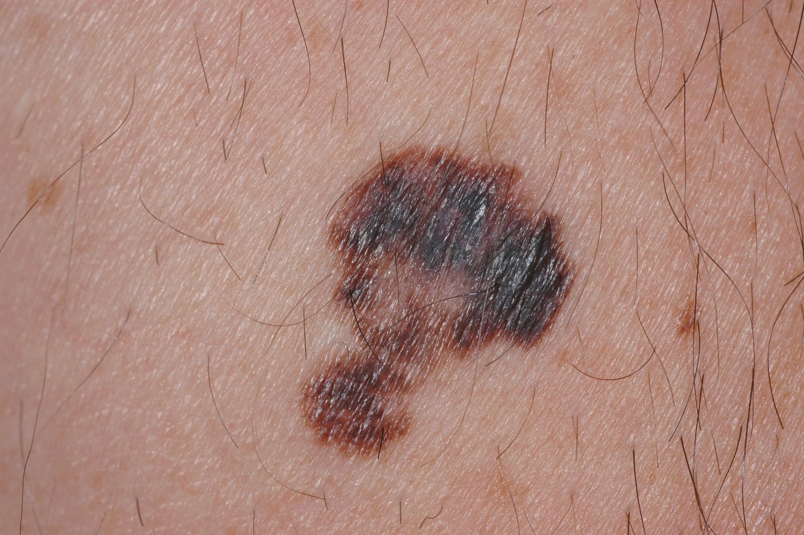
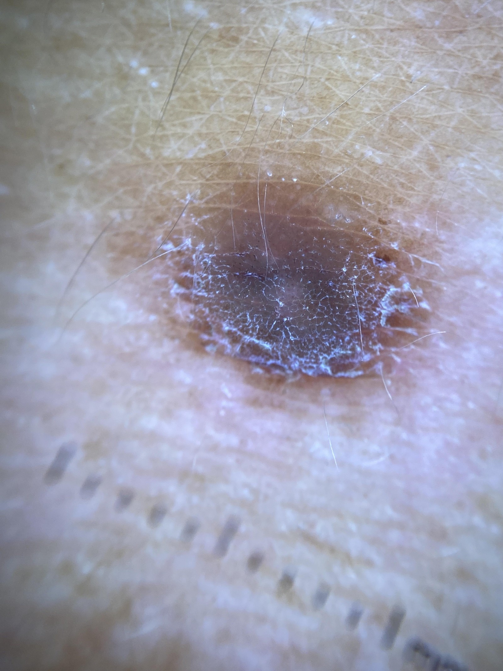
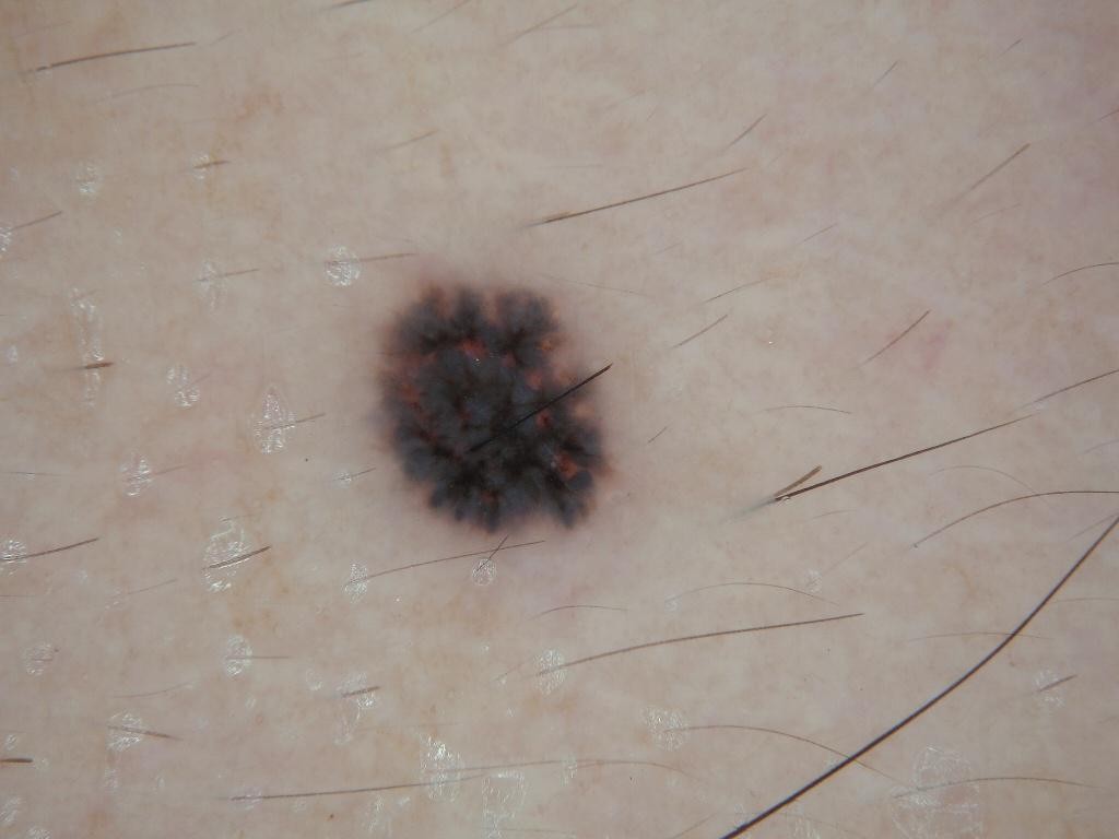
A 45-year-old man has the lesion shown, which his partner says has grown over the past few months. It is on his back and measures about 14 mm. What is the most appropriate next step?
Anonymous contributor, ISIC Archive ISIC_0024258, CC0 - ANarrow excisional biopsy with 1-3 mm clinical margins
- BReassure and re-photograph in 3 months
- CSuperficial tangential shave removal for cosmesis
- DCryotherapy with liquid nitrogen
- EWide local excision with 2 cm margins before any histology
Show answer
Answer A
Source AAD Guidelines of care for the management of primary cutaneous melanoma (Swetter SM et al., JAAD 2019)
- DR-003Multiple choiceDermatologyAdvanced
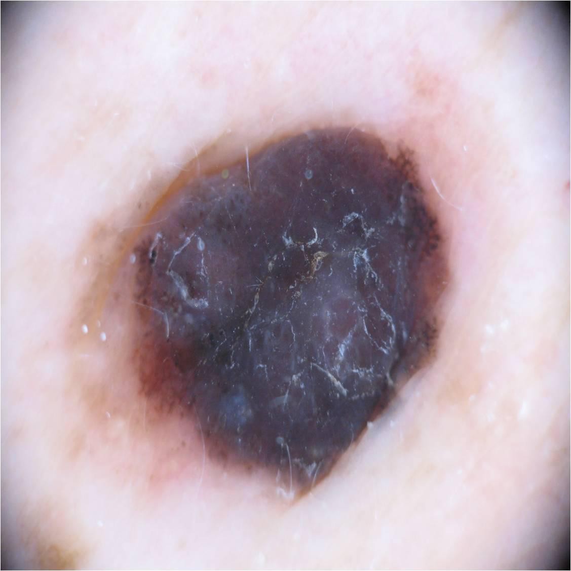
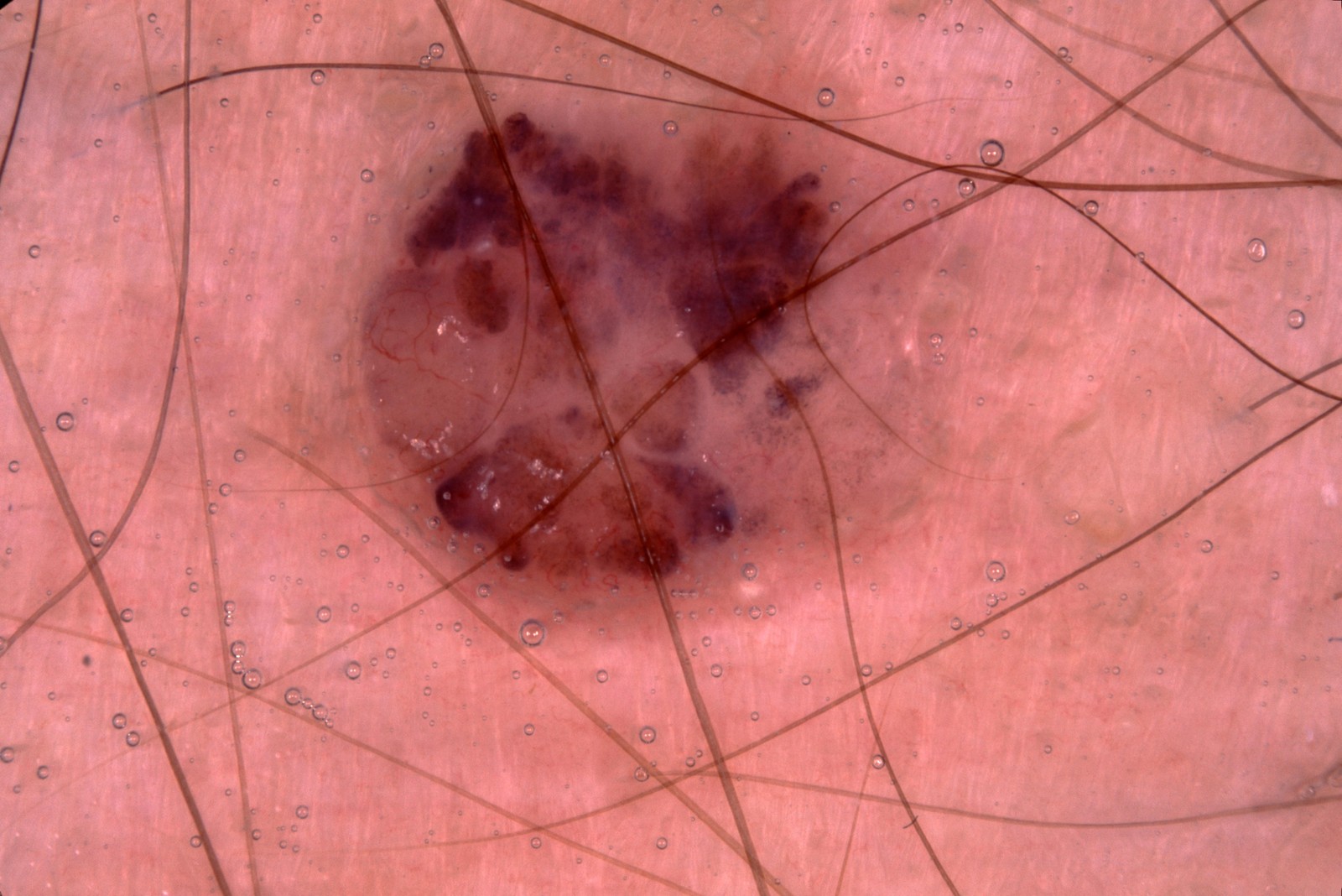
A 55-year-old woman noticed a raised dark lesion that has grown quickly over 4 months. The dermoscopic image is shown. What is the most likely diagnosis?
Anonymous contributor, ISIC Archive ISIC_0046450, CC BY - ABlue nevus
- BThrombosed hemangioma
- CPigmented basal cell carcinoma
- DSeborrheic keratosis
- ENodular melanoma
Show answer
Answer E
Source Kittler H et al. Standardization of terminology in dermoscopy/dermatoscopy, JAAD 2016; ISIC Archive histopathology label
- DR-004Multiple choiceDermatologyAdvanced
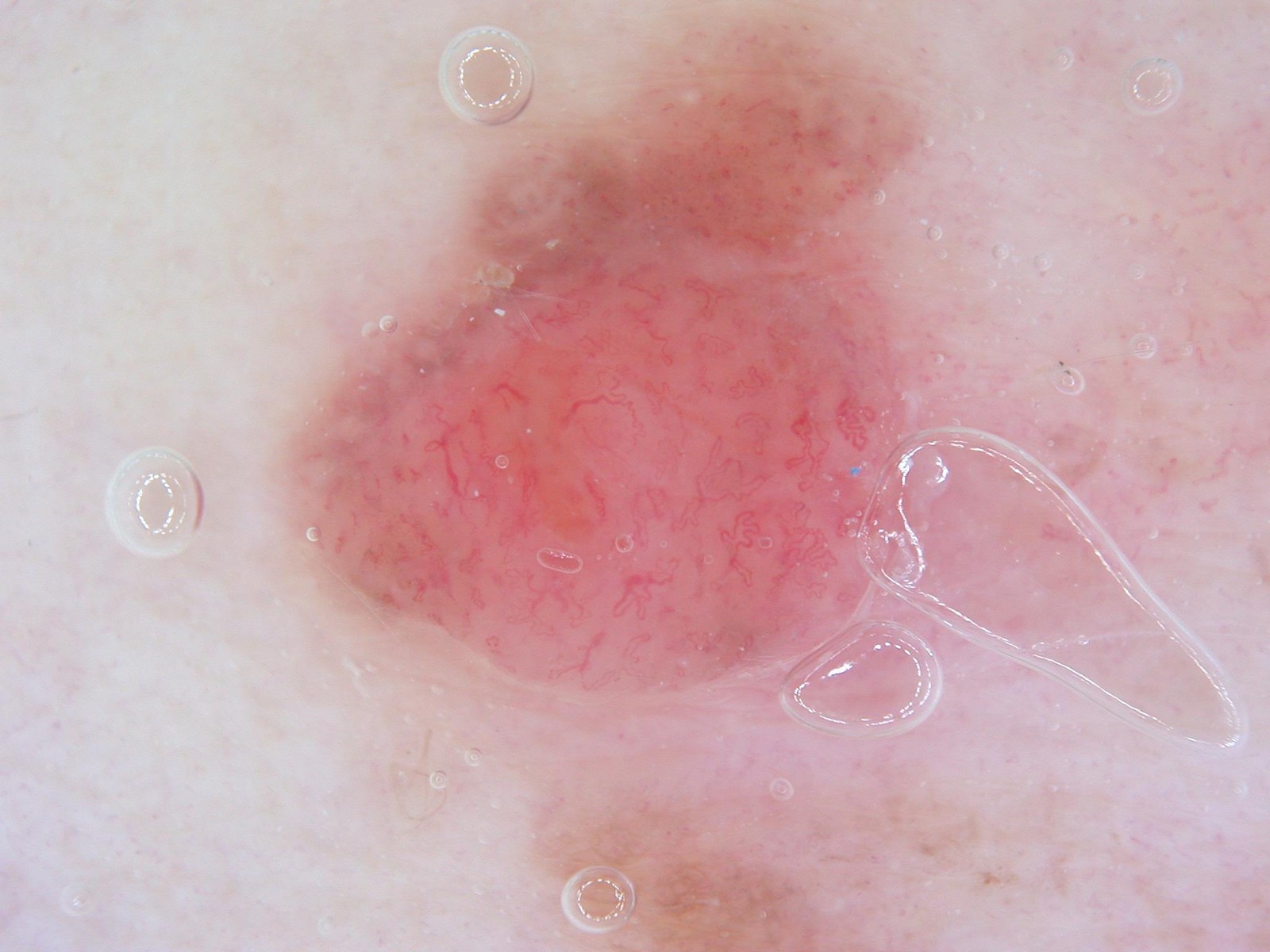
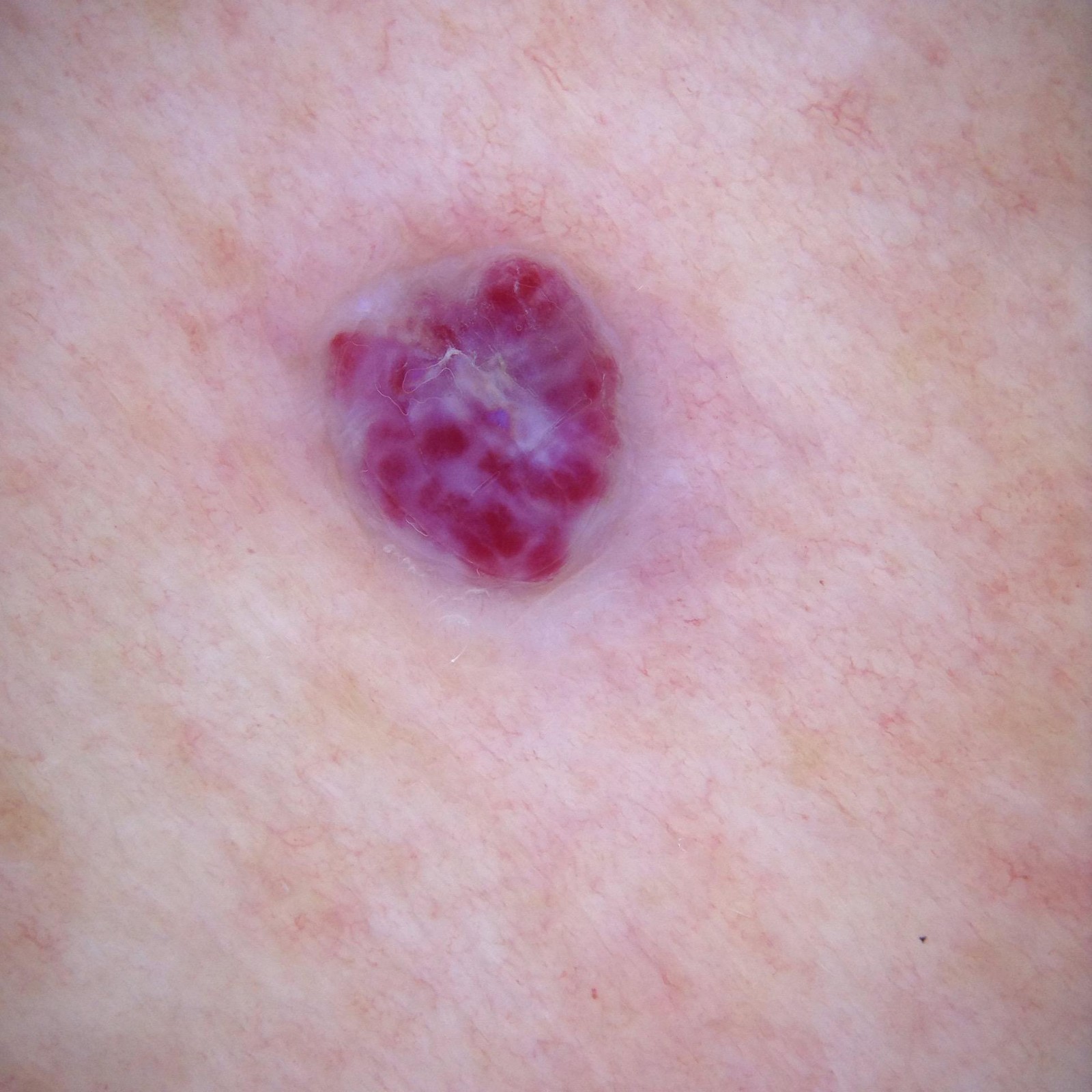
A 65-year-old woman has a pink papule that appeared and enlarged over 3 months. It is not painful and has not bled. The dermoscopic image is shown. What is the most likely diagnosis?
Anonymous contributor, ISIC Archive ISIC_0046626, CC BY - APyogenic granuloma
- BAmelanotic melanoma
- CNodular basal cell carcinoma
- DDermatofibroma
- EIntradermal (dermal) nevus
Show answer
Answer B
Source Kittler H et al. Standardization of terminology in dermoscopy/dermatoscopy, JAAD 2016; ISIC Archive histopathology label
- DR-005Multiple choiceDermatologyAdvanced
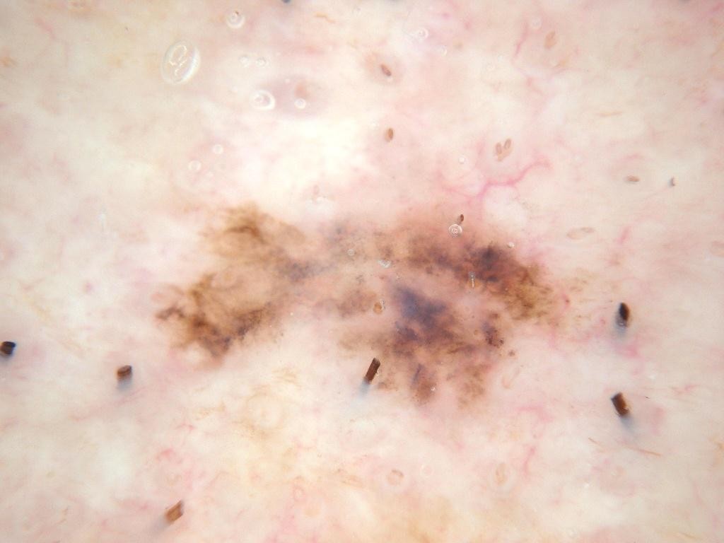
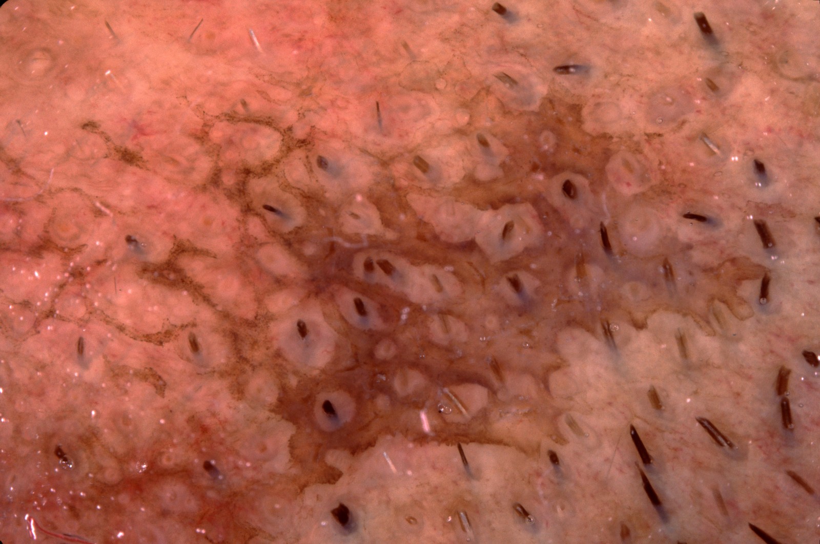
A 75-year-old man with extensive sun damage has a flat, irregular brown-gray patch on the sun-damaged skin of his head that has slowly enlarged over several years. The dermoscopic image is shown. What is the most likely diagnosis?
Anonymous contributor, ISIC Archive ISIC_0010067, CC0 - ASolar lentigo
- BPigmented actinic keratosis
- CFlat seborrheic keratosis
- DLentigo maligna (melanoma in situ)
- ELichen planus-like keratosis
Show answer
Answer D
Source Kittler H et al. Standardization of terminology in dermoscopy/dermatoscopy, JAAD 2016; ISIC Archive histopathology label
- DR-006Multiple choiceDermatologyBasic
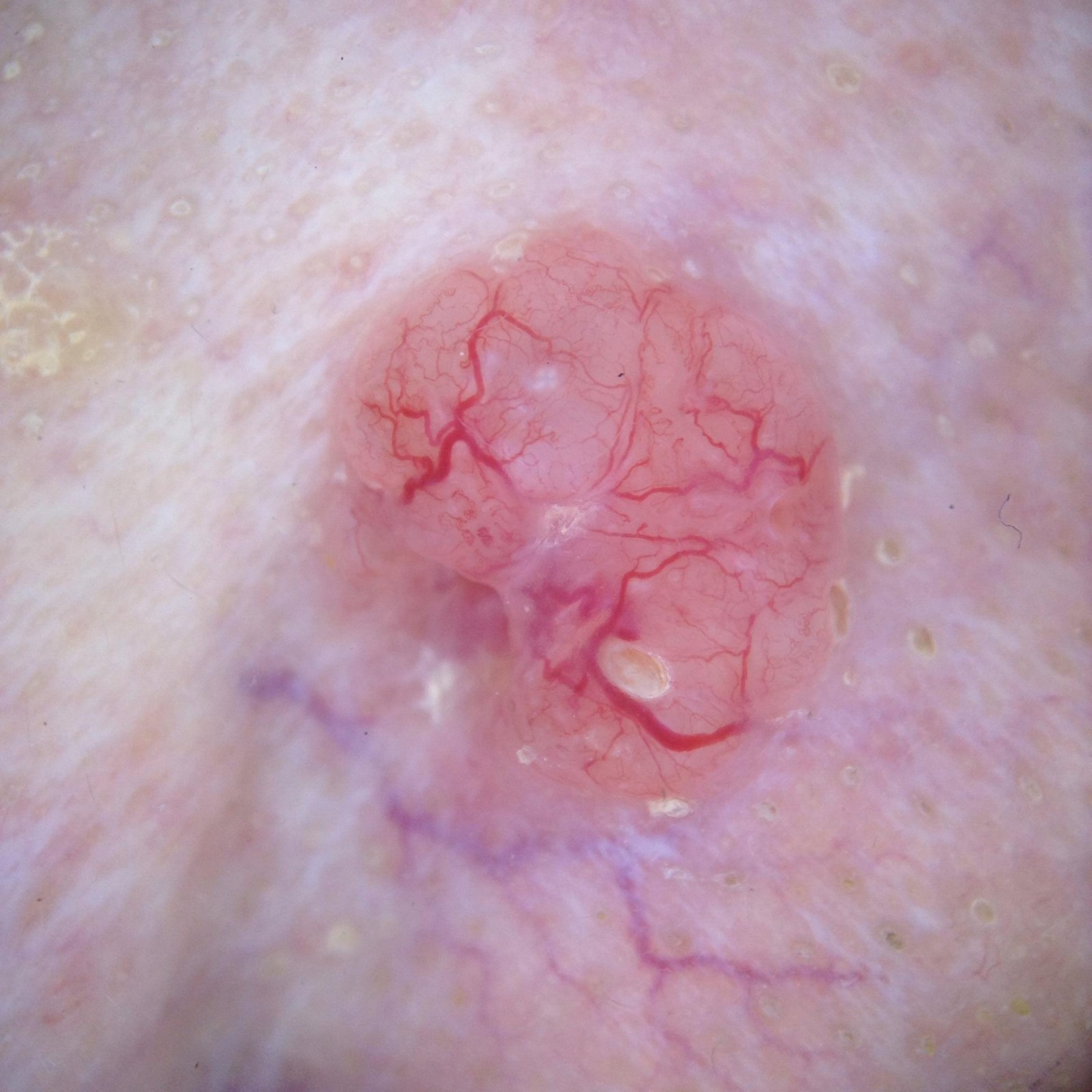
An 80-year-old woman has a slowly growing, shiny papule on her face. The dermoscopic image is shown. What is the most likely diagnosis?
Anonymous contributor, ISIC Archive ISIC_0001159, CC0 - ANodular basal cell carcinoma
- BSebaceous hyperplasia
- CAmelanotic melanoma
- DMolluscum contagiosum
- EIntradermal (dermal) nevus
Show answer
Answer A
Source Kittler H et al. Standardization of terminology in dermoscopy/dermatoscopy, JAAD 2016; ISIC Archive histopathology label
- DR-007Multiple choiceDermatologyIntermediate
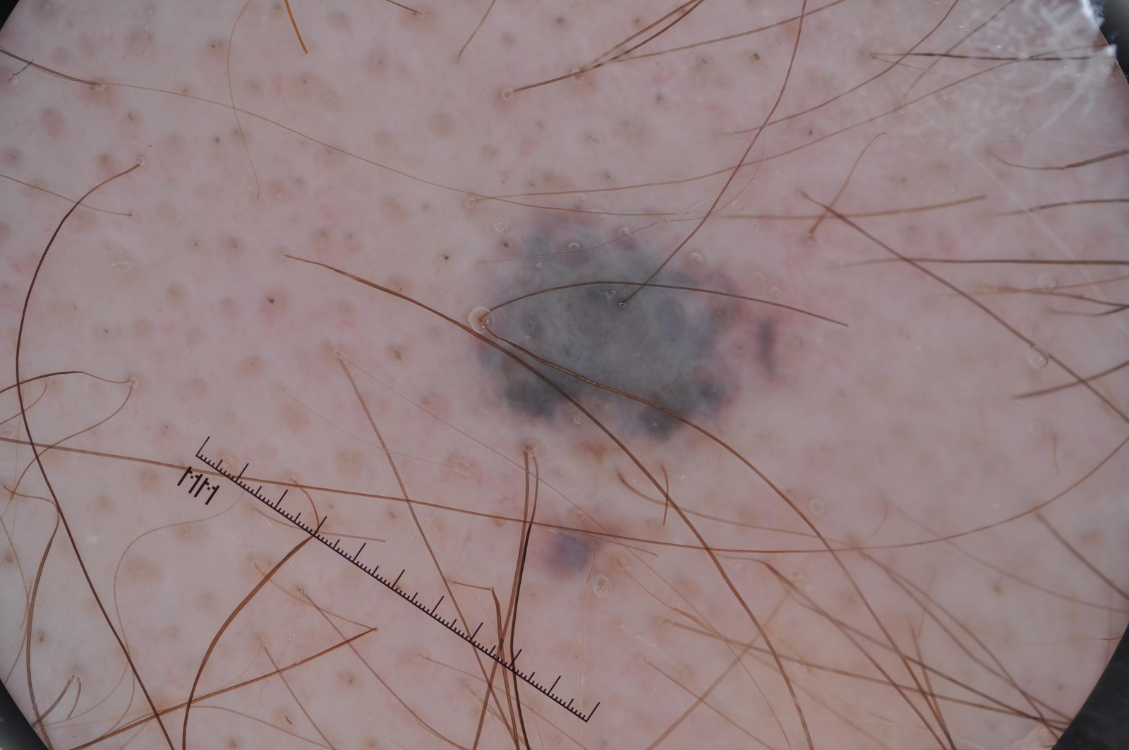
A 70-year-old man has a pigmented lesion on his upper back that his wife noticed a year ago. The dermoscopic image is shown. What is the most likely diagnosis?
Anonymous contributor, ISIC Archive ISIC_0011626, CC0 - AMelanoma
- BSeborrheic keratosis
- CBlue nevus
- DPigmented basal cell carcinoma
- EAngiokeratoma
Show answer
Answer D
Source Kittler H et al. Standardization of terminology in dermoscopy/dermatoscopy, JAAD 2016; ISIC Archive histopathology label
- DR-008Multiple choiceDermatologyIntermediate
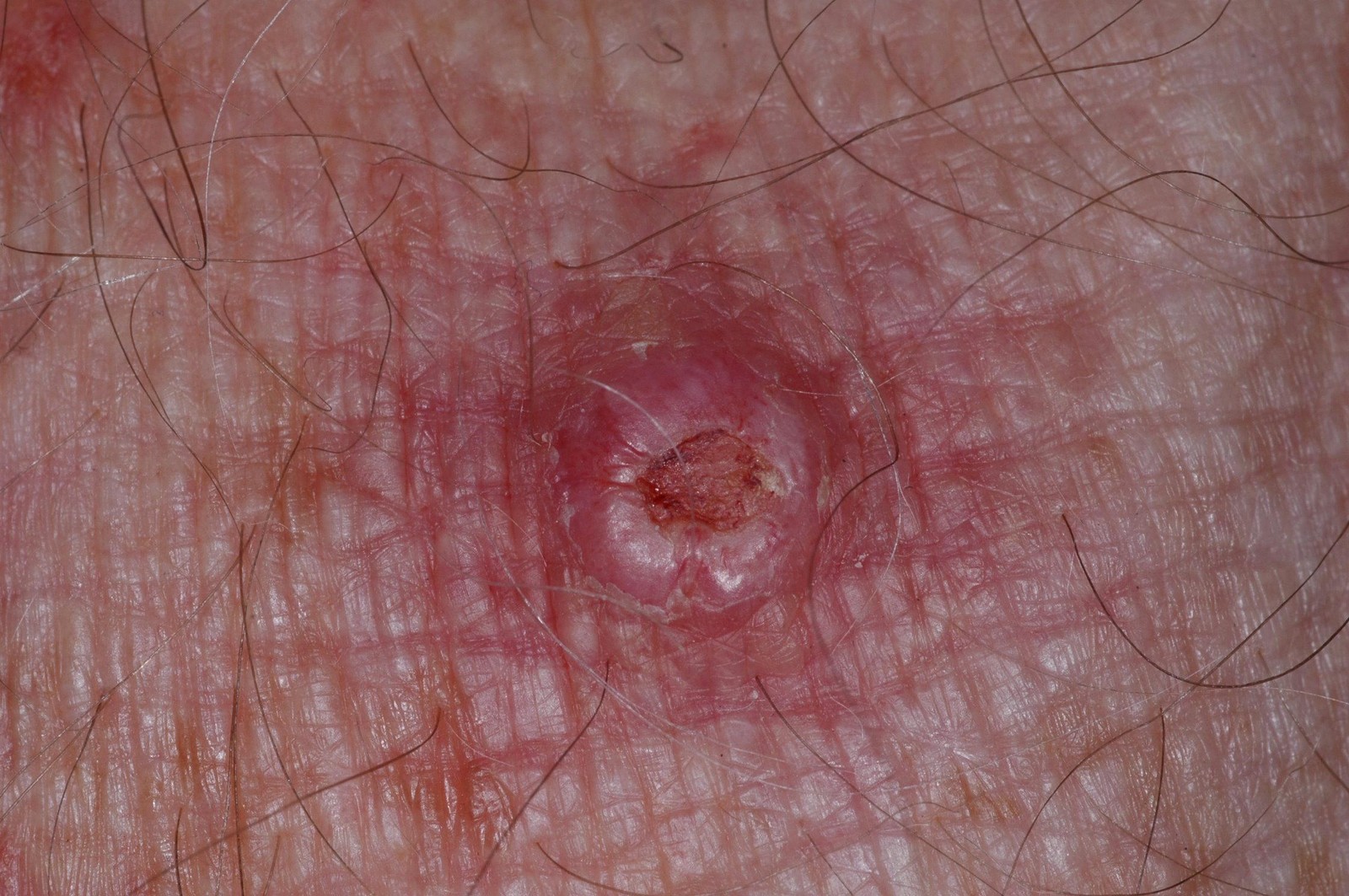
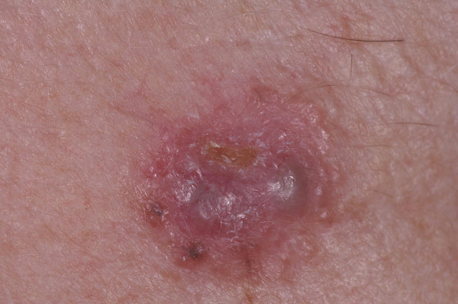
An 85-year-old man with heavily sun-damaged skin has the firm, tender, keratotic nodule shown, with a central crust, which has grown over a few months. What is the most likely diagnosis?
Anonymous contributor, ISIC Archive ISIC_0024212, CC0 - ANodular basal cell carcinoma
- BCutaneous squamous cell carcinoma
- CSeborrheic keratosis
- DAmelanotic melanoma
- EDermatofibroma
Show answer
Answer B
Source AAD Guidelines of care for the management of cutaneous squamous cell carcinoma (Alam M et al., JAAD 2018); ISIC Archive histopathology label
- DR-009Multiple choiceDermatologyIntermediate
A 75-year-old man has a waxy, raised brown lesion on his face, present for several years; he has similar lesions on his back. The dermoscopic image is shown. What is the most likely diagnosis?
Anonymous contributor, ISIC Archive ISIC_0010389, CC0 - AMelanoma
- BPigmented basal cell carcinoma
- CSolar lentigo
- DCongenital nevus
- ESeborrheic keratosis
Show answer
Answer E
Source Kittler H et al. Standardization of terminology in dermoscopy/dermatoscopy, JAAD 2016; ISIC Archive histopathology label
- DR-010Multiple choiceDermatologyBasic
An 85-year-old patient has a small red-purple papule that has been present for years. The dermoscopic image is shown. What is the most likely diagnosis?
Anonymous contributor, ISIC Archive ISIC_0001115, CC0 - ANodular melanoma
- BPyogenic granuloma
- CCherry hemangioma
- DKaposi sarcoma
- ENodular basal cell carcinoma
Show answer
Answer C
Source Kittler H et al. Standardization of terminology in dermoscopy/dermatoscopy, JAAD 2016; ISIC Archive histopathology label
- DR-011Multiple choiceDermatologyIntermediate
A 40-year-old woman has a firm, asymptomatic papule on her lower leg. The dermoscopic image is shown. What is the most likely diagnosis?
Hospital Italiano de Buenos Aires, ISIC Archive ISIC_5727659, CC BY - ADermatofibroma
- BMelanoma
- CBlue nevus
- DPigmented basal cell carcinoma
- ESeborrheic keratosis
Show answer
Answer A
Source Kittler H et al. Standardization of terminology in dermoscopy/dermatoscopy, JAAD 2016; ISIC Archive histopathology label
- DR-012Multiple choiceDermatologyIntermediate
A 55-year-old man has a small, flat blue-gray lesion that appears unchanged on photographs taken 20 years ago. The dermoscopic image is shown. What is the most likely diagnosis?
Anonymous contributor, ISIC Archive ISIC_0012489, CC0 - ANodular melanoma
- BThrombosed hemangioma
- CPigmented basal cell carcinoma
- DBlue nevus
- ESeborrheic keratosis
Show answer
Answer D
Source Kittler H et al. Standardization of terminology in dermoscopy/dermatoscopy, JAAD 2016; ISIC Archive histopathology label
- DR-013Multiple choiceDermatologyAdvanced
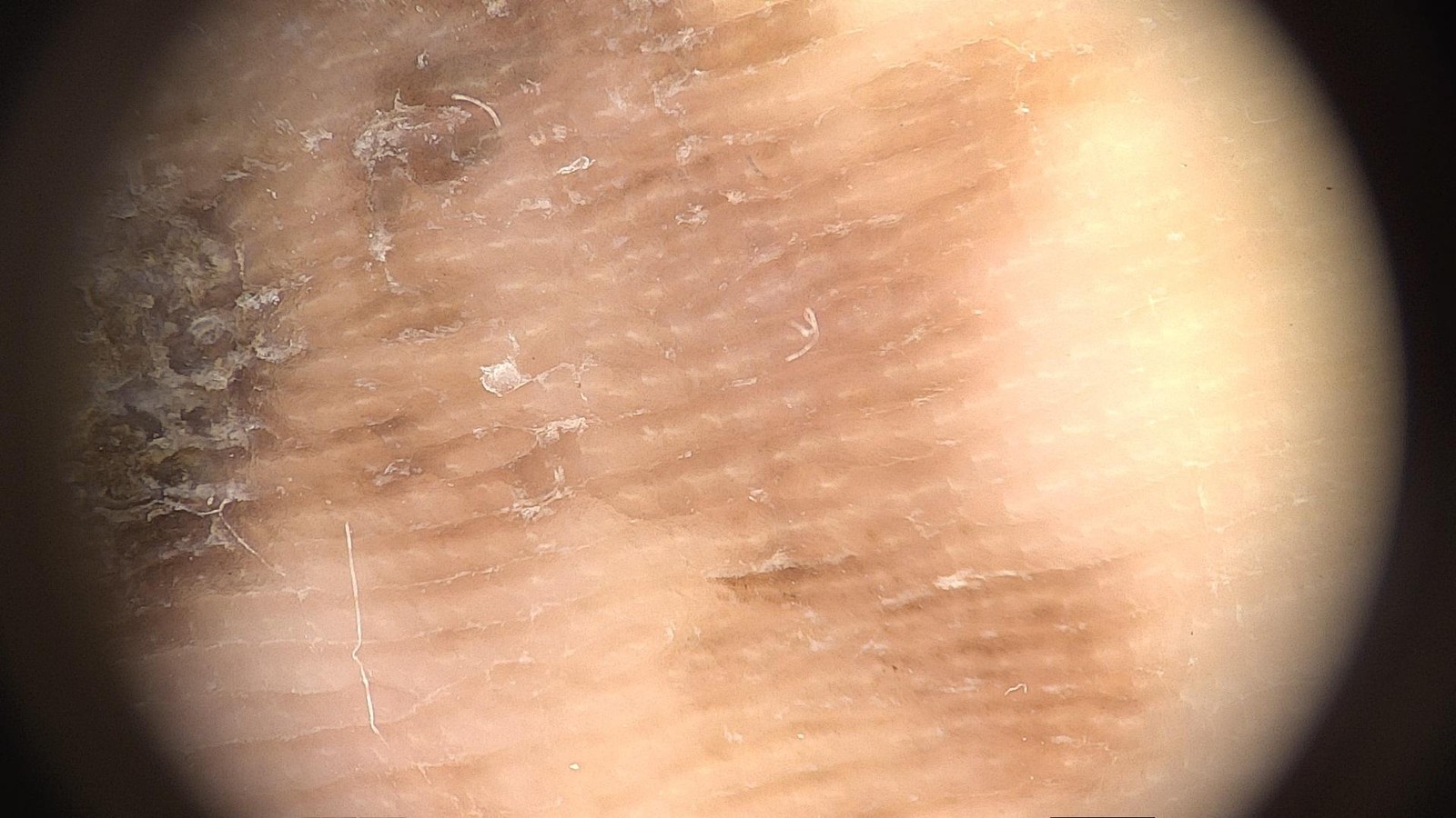
A 60-year-old woman has an irregular brown macule on the sole of her foot that has slowly enlarged over 2 years. The dermoscopic image is shown. What is the most likely diagnosis?
Department of Dermatology, University of Athens, Andreas Syggros Hospital of Skin and Venereal Diseases, Alexander Stratigos, Konstantinos Liopyris, ISIC Archive ISIC_3937563, CC BY - AAcral junctional nevus
- BAcral lentiginous melanoma in situ
- CSubcorneal hemorrhage (talon noir)
- DTinea nigra
- EPlantar wart
Show answer
Answer B
Source Kittler H et al. Standardization of terminology in dermoscopy/dermatoscopy, JAAD 2016; ISIC Archive histopathology label; Saida T et al., dermoscopy of acral pigmented lesions (parallel ridge pattern)
- DR-014Multiple choiceDermatologyIntermediate
An 85-year-old with a lifetime of outdoor work has a rough, scaly, pink-brown patch on the sun-exposed scalp that feels like sandpaper. The dermoscopic image is shown. What is the most likely diagnosis?
Anonymous contributor, ISIC Archive ISIC_0011552, CC0 - ALentigo maligna
- BSuperficial basal cell carcinoma
- CSeborrheic keratosis
- DDiscoid lupus erythematosus
- EActinic keratosis
Show answer
Answer E
Source Kittler H et al. Standardization of terminology in dermoscopy/dermatoscopy, JAAD 2016; ISIC Archive histopathology label
- DR-015Short answerDermatologyIntermediate
A 70-year-old woman has the slowly growing lesion shown, about 10 mm across, on her upper back. It bleeds occasionally. Give the most likely diagnosis, the clinical features in the image that support it, and how you would confirm and treat it.
Anonymous contributor, ISIC Archive ISIC_0024221, CC0 Rubric
- +3Diagnoses basal cell carcinoma (nodular)
- +2Names supporting features: pearly/translucent surface, rolled border, telangiectasias, central crust or ulceration
- +2Confirms with biopsy (shave or punch) before definitive treatment
- +2For a low-risk trunk lesion, recommends standard excision with about 4 mm margins or curettage and electrodesiccation
- -2Treats empirically with cryotherapy or a topical agent without histologic confirmation
- -2Calls it melanoma or a benign lesion and plans no treatment
Show answer
Reference answer Nodular basal cell carcinoma. Supporting features: a pink, translucent ('pearly') papule-nodule with a raised rolled border, fine telangiectasias over the surface, and a central crust or erosion. Confirm with a shave or punch biopsy. On the trunk, under 2 cm, primary and well defined, it is low risk: standard surgical excision with about 4 mm clinical margins and margin assessment, or curettage and electrodesiccation, are first-line. Mohs surgery is for high-risk tumors; radiotherapy or other options are for patients who cannot have surgery.
Source AAD Guidelines of care for the management of basal cell carcinoma (Kim JYS et al., JAAD 2018)
- DR-016Short answerDermatologyIntermediate
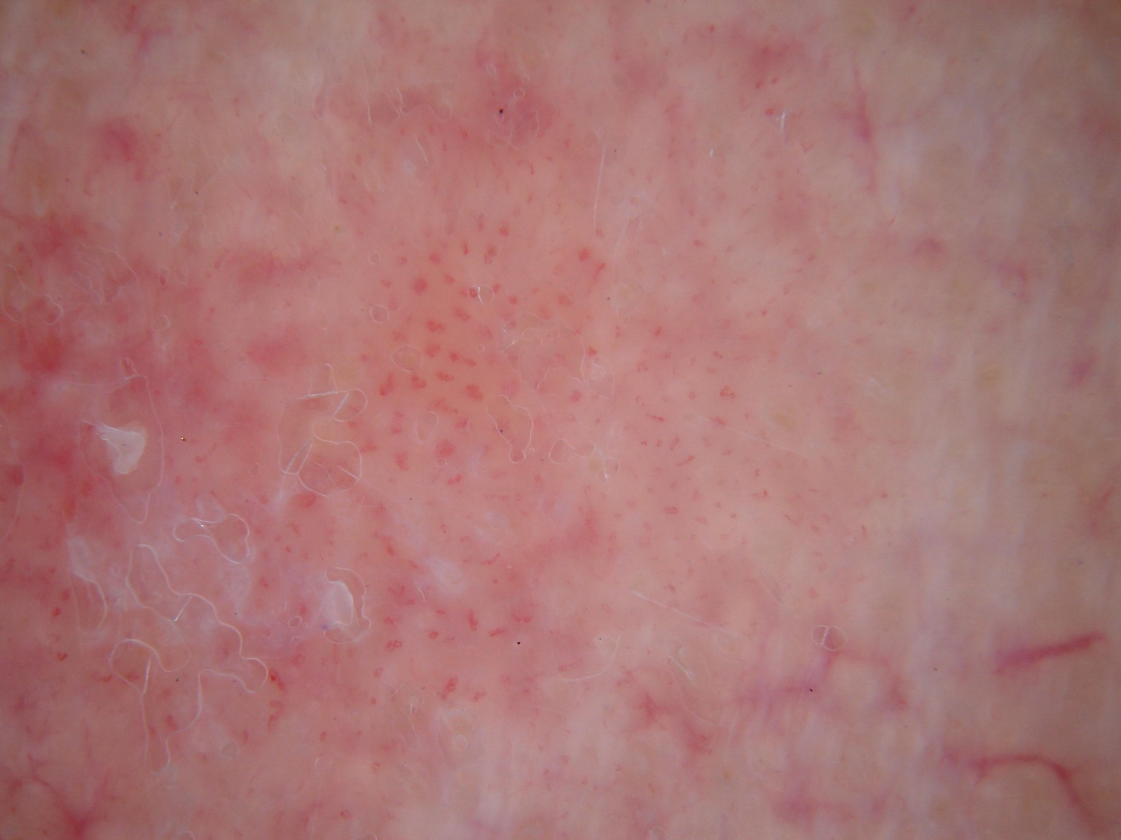
An 80-year-old man has a slowly enlarging, scaly, pink plaque on his scalp. The dermoscopic image is shown. Give the most likely diagnosis, the dermoscopic features that support it, and the management options.
Anonymous contributor, ISIC Archive ISIC_0020980, CC0 Rubric
- +3Diagnoses squamous cell carcinoma in situ (Bowen disease)
- +2Describes dotted or glomerular vessels on a pink background
- +1Notes surface scale
- +2Recommends biopsy to confirm and exclude invasive SCC or superficial BCC
- +2Lists at least two appropriate treatments (excision, curettage, cryotherapy, 5-FU, imiquimod, photodynamic therapy)
- -2Diagnoses psoriasis or eczema and treats with a topical steroid alone
Show answer
Reference answer Squamous cell carcinoma in situ (Bowen disease). Dermoscopy shows dotted and short coiled (glomerular-type) vessels, denser in places, on a pink background, with white-yellow surface scale. Psoriasis, superficial BCC and actinic keratosis are the main mimics, so confirm with biopsy. Treatment options include surgical excision, curettage and cautery, cryotherapy, topical 5-fluorouracil or imiquimod, and photodynamic therapy, chosen by size, site, healing potential and patient preference.
Source British Association of Dermatologists guidelines for the management of squamous cell carcinoma in situ (Bowen's disease) (Morton CA et al., Br J Dermatol 2014); Kittler H et al. Standardization of terminology in dermoscopy/dermatoscopy, JAAD 2016; ISIC Archive histopathology label
- DR-017Short answerDermatologyAdvanced
A 25-year-old man has a dark papule on his neck that appeared over the past year. The dermoscopic image is shown. What type of lesion is this, what important differential must be excluded, and how should it be managed?
Anonymous contributor, ISIC Archive ISIC_0011179, CC0 Rubric
- +3Identifies a spitzoid lesion (Spitz or Reed nevus)
- +2Describes peripheral clods and/or pseudopods/streaks around a dark (blue-black) center
- +2Names spitzoid melanoma as the differential to exclude
- +3Recommends complete excision for histopathology given the patient's adult age
- +1Suggests expert dermatopathology review
- -3Reassures without excision or follow-up
- -2Calls it a seborrheic keratosis or basal cell carcinoma
Show answer
Reference answer A spitzoid melanocytic lesion (Spitz/Reed nevus). Dermoscopy shows a dark lesion with a blue-gray to black center, scattered red-brown dots, and a rim of peripheral clods with short pseudopod-like projections (pigmented Spitz/Reed pattern, globular to starburst); there is some asymmetry. The key differential is spitzoid melanoma, which dermoscopy cannot reliably exclude. In patients aged 12 or older, spitzoid lesions should be completely excised for histopathology, ideally reviewed by an experienced dermatopathologist because atypical Spitz tumors exist.
Source Lallas A et al. The dermoscopic universe of Spitz/Reed nevi and management of Spitzoid lesions: an International Dermoscopy Society consensus (JAMA Dermatol 2017); Kittler H et al. Standardization of terminology in dermoscopy/dermatoscopy, JAAD 2016; ISIC Archive histopathology label
- DR-018Short answerDermatologyAdvanced
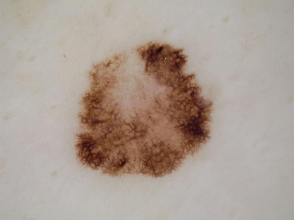
A 60-year-old woman has a pigmented lesion on her arm that she thinks has changed over the past 6 months. The dermoscopic image is shown. Which features concern you, what is your working diagnosis, and how would you manage it, including definitive surgical margins if histology shows melanoma in situ?
Anonymous contributor, ISIC Archive ISIC_0009882, CC0 Rubric
- +2Names at least two melanoma-specific structures (atypical network, irregular dots/globules, blotches, regression, asymmetry)
- +2Gives a working diagnosis of melanoma (in situ likely)
- +2Recommends narrow excisional biopsy with 1-3 mm margins
- +2States 0.5-1 cm margins for melanoma in situ
- +1States that sentinel node biopsy is not indicated for melanoma in situ
- -3Recommends reassurance or monitoring only
- -2Recommends 2 cm margins or sentinel node biopsy for melanoma in situ
Show answer
Reference answer Concerning features: asymmetry of color and structure, an atypical pigment network with thickened, irregular lines, irregular dark dots, globules and blotches at the periphery, and a central structureless hypopigmented area suggesting regression. Working diagnosis: melanoma, probably in situ; a dysplastic nevus is the main alternative. Do a narrow excisional biopsy with 1-3 mm margins. If it is melanoma in situ, re-excise with 0.5-1 cm clinical margins. Sentinel node biopsy and staging imaging are not indicated for in situ disease; arrange a full skin examination and follow-up.
Source AAD Guidelines of care for the management of primary cutaneous melanoma (Swetter SM et al., JAAD 2019)
- DR-019Short answerDermatologyIntermediate
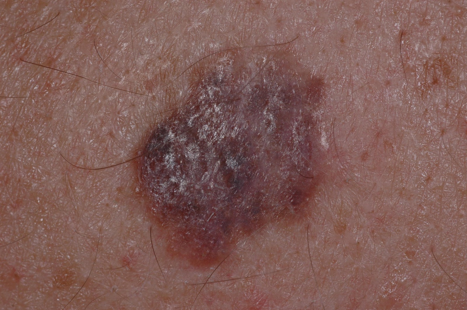
A 40-year-old man has the lesion shown (about 15 mm) on his back, which has changed over the past year. (a) What is the most likely diagnosis, and which features in the image support it? (b) An excisional biopsy confirms invasive melanoma, Breslow thickness 0.8 mm, not ulcerated, margins clear. What definitive surgery do you recommend, and what about sentinel lymph node biopsy?
Anonymous contributor, ISIC Archive ISIC_0024298, CC0 Rubric
- +2Diagnoses melanoma
- +2Names supporting features: asymmetry, irregular border, multiple colors, size over 6 mm
- +3Recommends wide local excision with 1 cm margins
- +2States sentinel node biopsy should be discussed/considered for a T1b (0.8-1.0 mm) melanoma
- +1Recommends ongoing full skin examination and follow-up
- -3Recommends 2 cm margins or routine CT/PET staging
- -2States that sentinel node biopsy is never relevant at this thickness
Show answer
Reference answer (a) Melanoma: an asymmetric lesion larger than 6 mm with an irregular border and several colors (brown, violaceous, blue-gray and pink), an irregular surface and documented change over a year. (b) This is T1b (0.8-1.0 mm). Wide local excision with 1 cm clinical margins. Sentinel lymph node biopsy should be discussed and can be considered for T1b tumors, which carry roughly a 5-10% chance of a positive node. Routine cross-sectional imaging is not indicated. Arrange full skin examinations and follow-up.
Source AAD Guidelines of care for the management of primary cutaneous melanoma (Swetter SM et al., JAAD 2019); NCCN Guidelines: Melanoma: Cutaneous (2024)