@joshua/ecg-interpretation-12-lead
ECG interpretation: 12-lead tracings
Author-made · not reviewedCardiologyCC BY 4.0
Written by the BenchClub team with AI help. Answers follow public guidelines and textbooks; not yet reviewed by a practising physician.
v117 questions17 with imagesUpdated Nov 17, 2026
- ECG-001Short answerCardiologyAdvanced
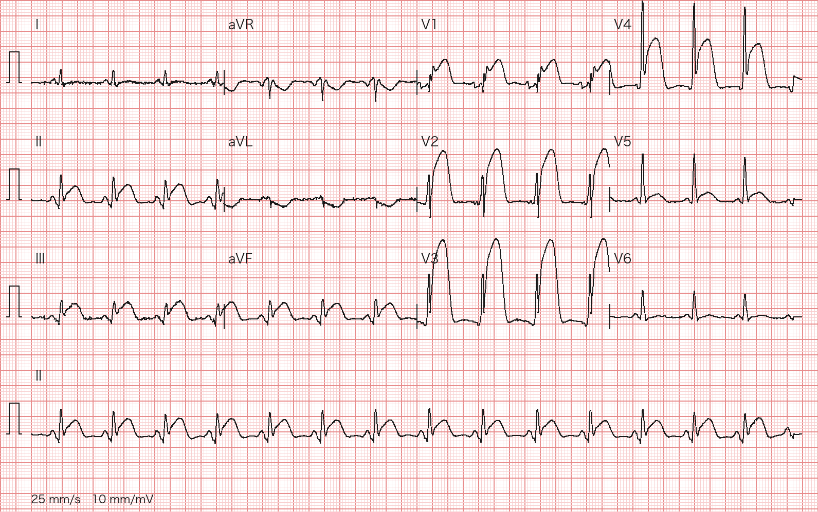
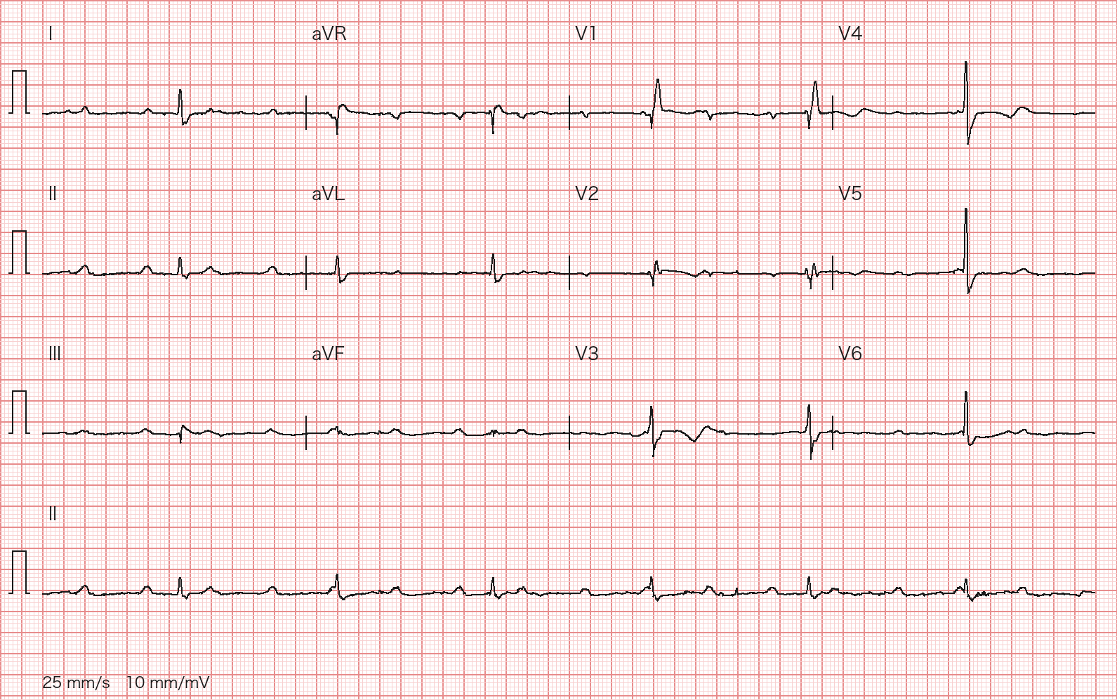
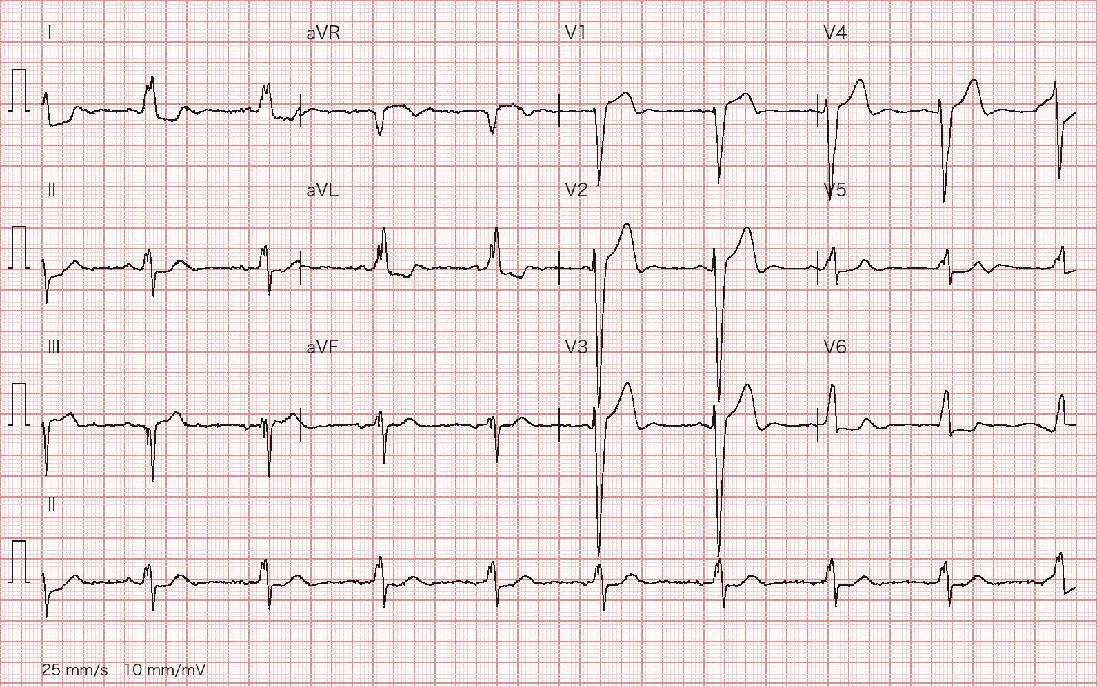
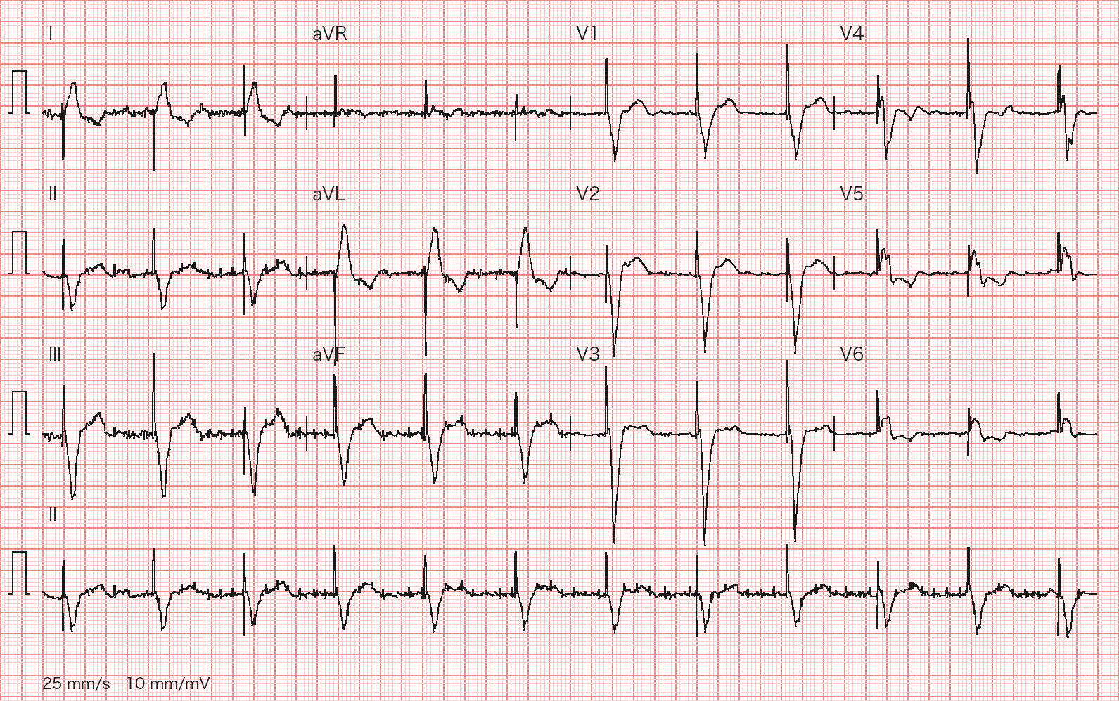
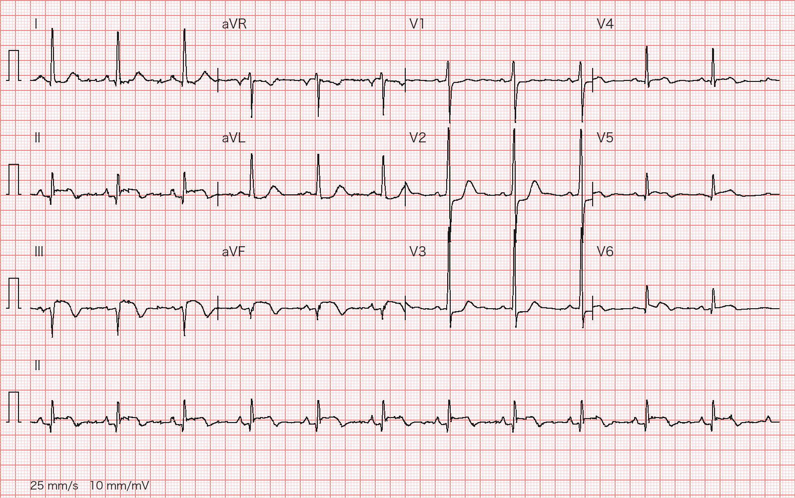
A 69-year-old man has had crushing substernal chest pain for 90 minutes with diaphoresis. BP 104/68 mmHg, HR 88 bpm, SpO₂ 97% on room air, lungs clear. The hospital offers 24/7 primary PCI. His first ED ECG is shown (12-lead ECG at 25 mm/s and 10 mm/mV; 10-second lead II rhythm strip at the bottom). Describe: (1) the key ECG findings and diagnosis; (2) the most likely culprit artery and why; (3) your management over the next 30 minutes.
Wagner P, Strodthoff N et al., PTB-XL 1.0.3, PhysioNet, CC BY 4.0; redrawn by BenchClub from the raw waveform Rubric
- +3Identifies precordial ST elevation (V1–V4 or V2–V5 acceptable) with acute anterior STEMI (2 points) and the additional inferior ST elevation (1 point)
- +2Names the LAD (wrap-around LAD) as the most likely culprit (1 point) and reasons about the level of occlusion from the ECG, either beyond D1 (no I/aVL elevation) or proximal (V1 elevation) (1 point)
- +3Activates primary PCI immediately without waiting for troponin
- +1Obtains V3R/V4R to assess RV involvement
- +1Gives aspirin loading and anticoagulation (UFH or bivalirudin); both required; P2Y12 timing per cath-lab protocol is acceptable
- -3Waits for troponin or a repeat ECG before deciding on reperfusion, or chooses fibrinolysis despite available primary PCI
- -2Gives nitrates with SBP <90 mmHg, recent PDE-5 inhibitor use, or suspected RV infarction
- -2Reads the tracing as acute pericarditis or early repolarization
Show answer
Reference answer (1) Sinus rhythm about 88 bpm, narrow QRS. Massive convex (tombstone) ST elevation in V2–V4 (maximal in V3), ST elevation in V1, modest ST elevation (about 1–3 mm at J+60 ms, most evident in III and aVF) in II, III and aVF, minimal in V5, none in V6, and no ST elevation in I or aVL. Diagnosis: acute anterior STEMI with inferior involvement. (2) Anterior plus inferior elevation without elevation in I/aVL points to a 'wrap-around' LAD that supplies the inferior apex; inferior elevation with none in I/aVL favors occlusion beyond the first diagonal, although prominent V1 elevation raises the possibility of a more proximal (pre-first-septal) lesion. Proximal RCA occlusion with RV infarction can also elevate V1, but then V1 elevation is usually ≥ V2 and fades across V2–V3; the reverse gradient here favors the LAD. Angiography decides. (3) Activate the cath lab immediately on ECG diagnosis without waiting for troponin (first-medical-contact-to-device ≤90 min). Record right-sided leads V3R/V4R now. Chewed aspirin 162–325 mg and IV unfractionated heparin; P2Y12 loading with ticagrelor 180 mg or prasugrel 60 mg (no prior stroke/TIA; clopidogrel 600 mg if both are contraindicated), in the ED or the lab per local protocol. Oxygen only if SpO₂ <90%. Continuous monitoring with defibrillator pads available. Sublingual nitroglycerin for ongoing pain is reasonable if SBP ≥90 mmHg (and not ≥30 mmHg below baseline), no PDE-5 inhibitor within 24–48 h, and no suspected RV infarction. Watch for cardiogenic shock and VF.
Source PTB-XL 1.0.3 cardiologist annotation (ecg_id 1063); 2025 ACC/AHA/ACEP/NAEMSP/SCAI Guideline for the Management of Patients With Acute Coronary Syndromes
- ECG-002Multiple choiceCardiologyIntermediate
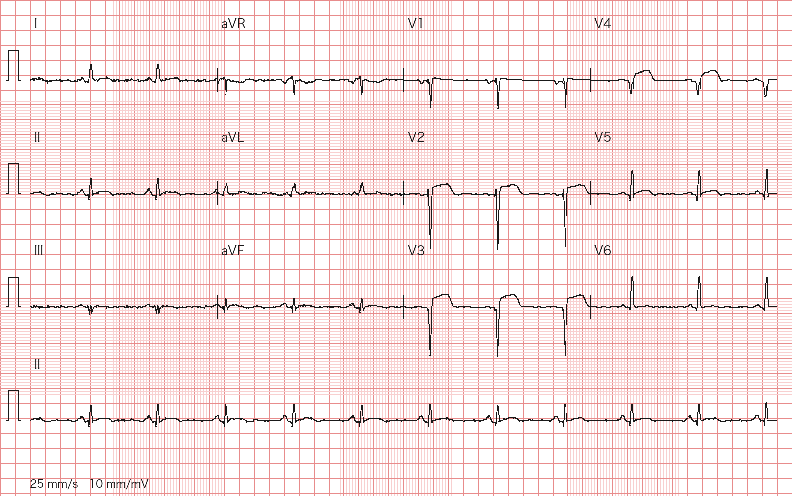
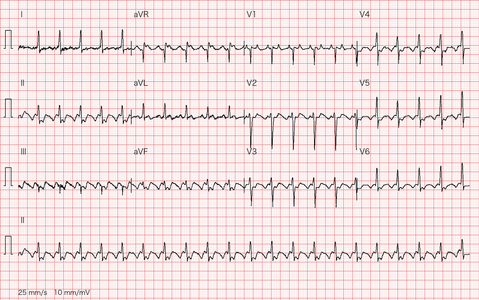
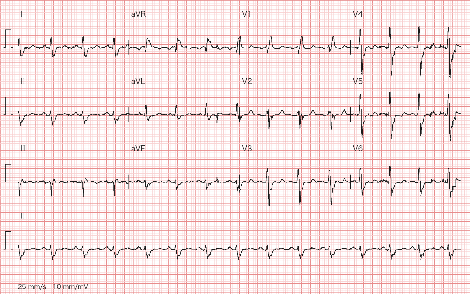
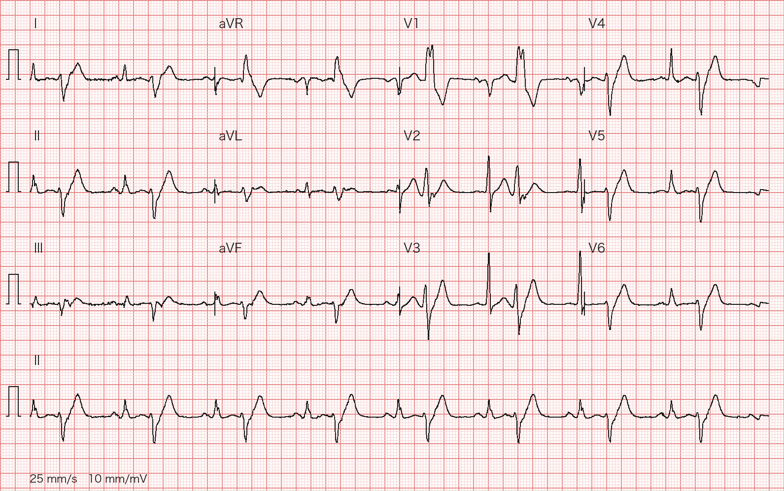
A 73-year-old man has had unrelenting chest pressure for 20 hours. BP 132/80 mmHg, hs-cTnT elevated. His ECG is shown (12-lead ECG at 25 mm/s and 10 mm/mV; 10-second lead II rhythm strip at the bottom). Which statement about infarct location and management is most appropriate?
Wagner P, Strodthoff N et al., PTB-XL 1.0.3, PhysioNet, CC BY 4.0; redrawn by BenchClub from the raw waveform - AAnterior STEMI, sinus tachycardia about 110 bpm; primary PCI
- BAnterior STEMI, sinus rhythm about 65 bpm; primary PCI
- CInferior STEMI, sinus rhythm about 65 bpm; primary PCI
- DAnterior STEMI with complete heart block (ventricular rate about 40 bpm); temporary pacing first, then PCI
- EAnterior STEMI beyond 12 hours; IV fibrinolysis is preferred
Show answer
Answer B
Source PTB-XL 1.0.3 cardiologist annotation (ecg_id 11353); 2025 ACC/AHA/ACEP/NAEMSP/SCAI Guideline for the Management of Patients With Acute Coronary Syndromes; 2023 ESC Guidelines for the management of acute coronary syndromes
- ECG-003Short answerCardiologyAdvanced
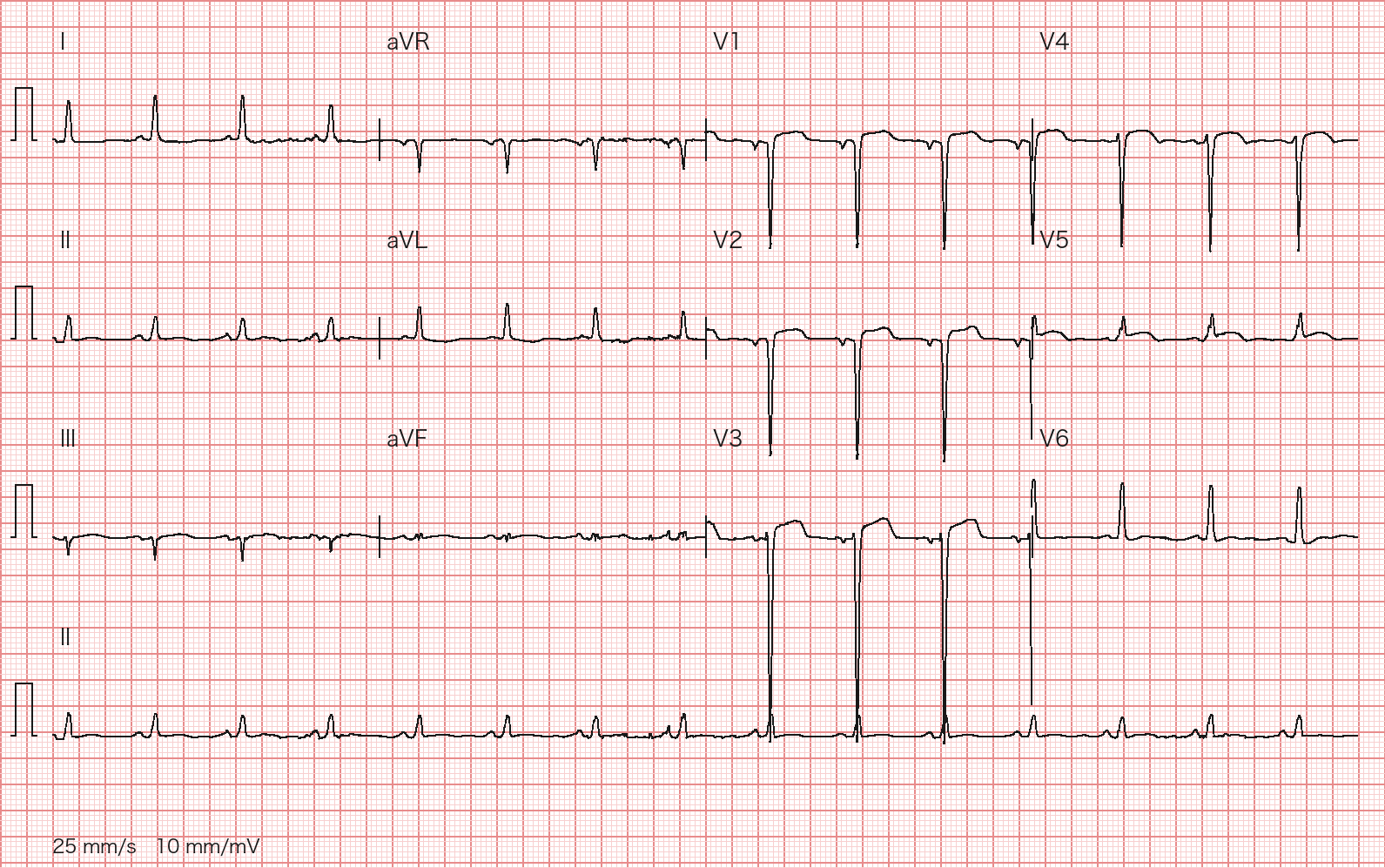
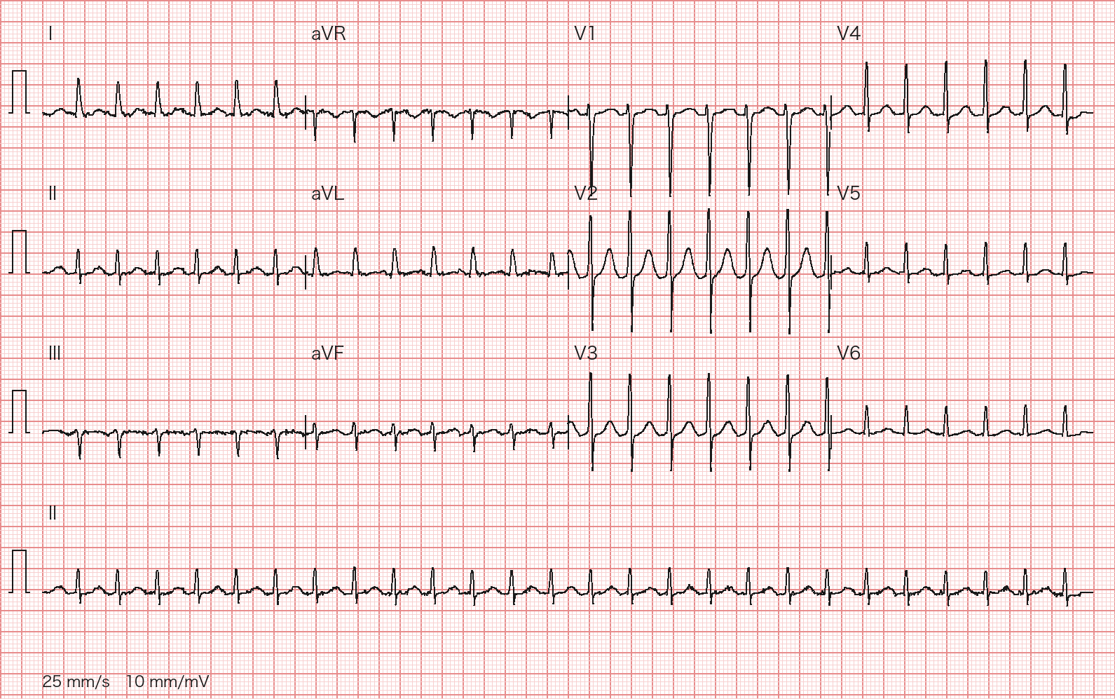
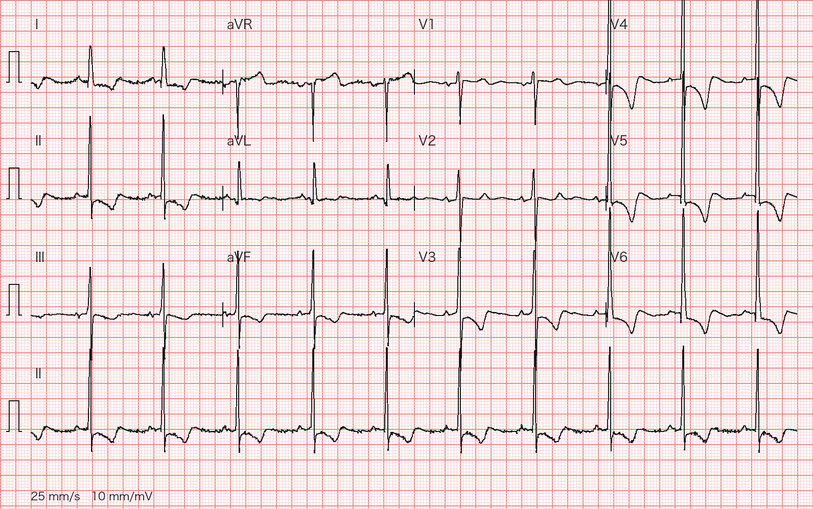
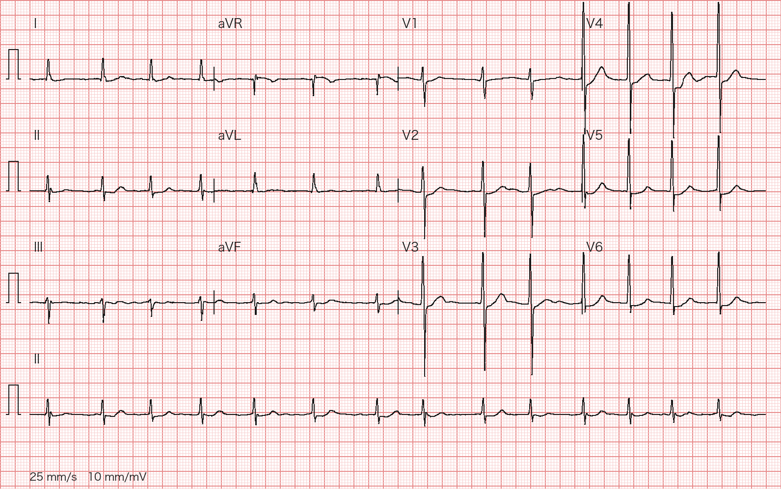
A 75-year-old woman was admitted elsewhere 8 months ago for an anterior MI and did not receive reperfusion. Over the past month she has had worsening exertional dyspnea and no chest pain. Two hs-troponins 3 hours apart are normal with no rise or fall. Her ECG is shown (12-lead ECG at 25 mm/s and 10 mm/mV; 10-second lead II rhythm strip at the bottom). Give the most likely interpretation, what must be excluded, and the next investigations.
Wagner P, Strodthoff N et al., PTB-XL 1.0.3, PhysioNet, CC BY 4.0; redrawn by BenchClub from the raw waveform Rubric
- +3Interprets as persistent ST elevation after an old anterior MI (LV aneurysm or dyskinetic segment)
- +1Notes anterior QS complexes (V1–V4; V1–V3 acceptable) with ST elevation
- +1Names at least one feature against acute STEMI (ST elevation small relative to QS depth, no hyperacute T waves, or no reciprocal depression)
- +1Cites the absence of chest pain and the flat troponins
- +1Compares with prior ECGs (or serial ECGs/troponins) to exclude recurrent MI
- +2Requests imaging for aneurysm, LVEF and mural thrombus (TTE; contrast echo or cardiac MRI preferred for thrombus)
- +1Any of: heart failure workup (e.g., NT-proBNP, GDMT if LVEF reduced), ischemia/viability evaluation, or anticoagulation if thrombus is found
- -3Calls it an acute STEMI and activates emergency PCI or fibrinolysis
Show answer
Reference answer Sinus rhythm about 90 bpm, normal axis (about 0° to +30°), narrow QRS; QS complexes in V1–V4 with about 1–3 mm ST elevation in V1–V4 (maximal in V3) that is small relative to the QS depth (low T/QRS ratio), without hyperacute T waves or reciprocal depression. With an anterior MI 8 months ago, no chest pain and flat troponins, this is most likely persistent ST elevation after an old anterior MI, suggesting LV aneurysm (or a dyskinetic segment), not a new STEMI. Recurrent acute anterior MI must be excluded: symptoms, serial ECGs and troponins, and comparison with prior tracings if they can be obtained. Next: transthoracic echo for aneurysm, LVEF and mural thrombus (contrast echo or cardiac MRI is more sensitive for thrombus); evaluate the dyspnea as heart failure (NT-proBNP; guideline-directed therapy if LVEF is reduced) and for ischemia/revascularization given unrevascularized CAD (angiography or CCTA, or ischemia/viability imaging such as CMR); anticoagulate if thrombus is found.
Source PTB-XL 1.0.3 cardiologist annotation (ecg_id 8636); Fourth Universal Definition of Myocardial Infarction (2018); 2025 ACC/AHA/ACEP/NAEMSP/SCAI Guideline for the Management of Patients With Acute Coronary Syndromes
- ECG-004Multiple choiceCardiologyBasic
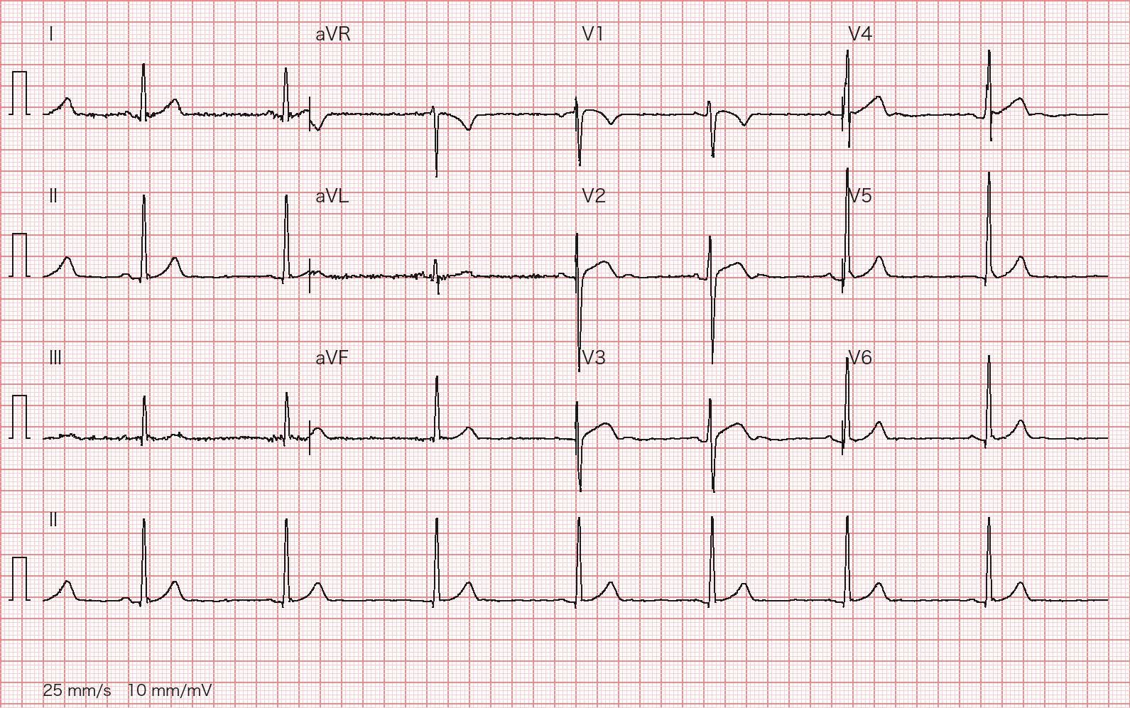
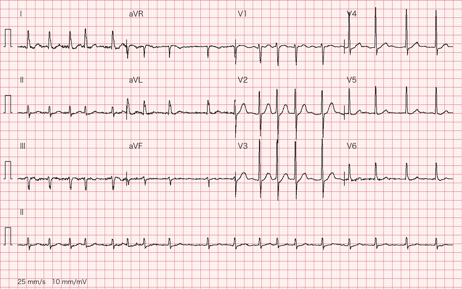
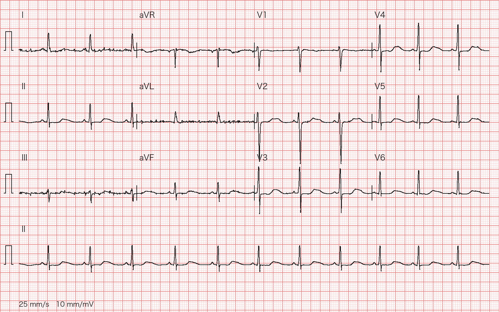
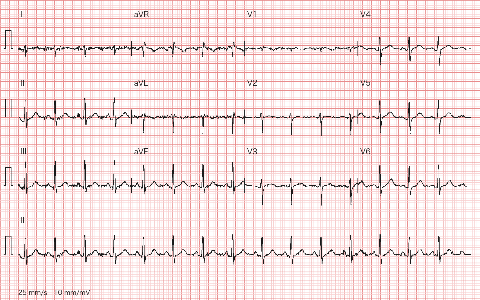
A 25-year-old man has a routine physical. He is asymptomatic, with no syncope and no family history of sudden death. His ECG is shown (12-lead ECG at 25 mm/s and 10 mm/mV; 10-second lead II rhythm strip at the bottom). Which description is most accurate?
Wagner P, Strodthoff N et al., PTB-XL 1.0.3, PhysioNet, CC BY 4.0; redrawn by BenchClub from the raw waveform - ASinus rhythm about 75 bpm with mild concave ST elevation in V2–V3, a normal variant
- BSinus bradycardia with first-degree AV block (PR >200 ms)
- CJunctional escape rhythm about 45 bpm
- DSecond-degree AV block, Mobitz type I
- ESinus bradycardia about 45 bpm with mild concave ST elevation in V2–V3, a normal variant
Show answer
Answer E
Source PTB-XL 1.0.3 cardiologist annotation (ecg_id 920); 2017 International Recommendations for Electrocardiographic Interpretation in Athletes
- ECG-005Short answerCardiologyIntermediate
An 88-year-old woman has had two syncopal episodes without prodrome in the past week. She takes no beta-blocker, non-dihydropyridine calcium channel blocker or digoxin. BP 150/60 mmHg, alert. Her ED ECG is shown (12-lead ECG at 25 mm/s and 10 mm/mV; 10-second lead II rhythm strip at the bottom). Give the ECG diagnosis and management.
Wagner P, Strodthoff N et al., PTB-XL 1.0.3, PhysioNet, CC BY 4.0; redrawn by BenchClub from the raw waveform Rubric
- +3Diagnoses third-degree (complete) AV block, noting P waves marching through a regular ~40 bpm escape with constant R-R; advanced/high-grade AV block with a ~40 bpm escape also earns full credit
- +1Identifies a subsidiary escape rhythm around 40 bpm (junctional or ventricular both acceptable)
- +2States that a permanent pacemaker is needed
- +2Evaluates for reversible causes (hyperkalemia/electrolytes, ischemia/inferior MI, hypothyroidism, offending drugs) without delaying pacing
- +1Continuous monitoring with transcutaneous or transvenous temporary pacing as a bridge if needed
- -3Recommends observation without pacing evaluation (e.g., after reading it as Mobitz I, 2:1 AV block or sinus bradycardia)
- -2Treats atropine as the main and sufficient therapy
Show answer
Reference answer P waves regular at about 100/min and QRS complexes regular at about 40/min with no fixed relationship. Because the rate ratio is about 2.5:1 the PR appears to alternate between two values, but the R-R stays constant and no QRS comes early after a well-timed P wave, so there is no conduction (AV dissociation). This is most consistent with third-degree (complete) AV block (at minimum high-grade AV block; a longer strip would confirm complete dissociation) with a subsidiary escape rhythm (junctional or ventricular; narrow-to-borderline QRS). Management: check potassium, cardiac biomarkers, thyroid function and other reversible causes; continuous monitoring with transcutaneous pacing pads applied; temporary pacing if hemodynamically unstable (atropine may be tried but is unreliable; isoproterenol or dopamine can bridge). Acquired complete heart block not due to a reversible cause is a Class 1 indication for permanent pacing (2018 ACC/AHA/HRS); with recurrent syncope it should be done promptly.
Source PTB-XL 1.0.3 cardiologist annotation (ecg_id 959); 2018 ACC/AHA/HRS Guideline on the Evaluation and Management of Bradycardia and Cardiac Conduction Delay
- ECG-006Multiple choiceCardiologyIntermediate
A 56-year-old man has had palpitations for 1 day. BP 128/80 mmHg, no chest pain. His ECG is shown (12-lead ECG at 25 mm/s and 10 mm/mV; 10-second lead II rhythm strip at the bottom). What is the most likely rhythm?
Wagner P, Strodthoff N et al., PTB-XL 1.0.3, PhysioNet, CC BY 4.0; redrawn by BenchClub from the raw waveform - ASinus tachycardia about 130 bpm
- BAV nodal reentrant tachycardia about 180 bpm
- CAtrial flutter with 2:1 AV conduction, ventricular rate about 130 bpm
- DAtrial flutter with 4:1 AV conduction, ventricular rate about 65 bpm
- EAtrial fibrillation with rapid ventricular response
Show answer
Answer C
Source PTB-XL 1.0.3 cardiologist annotation (ecg_id 23); 2015 ACC/AHA/HRS Guideline for the Management of Adult Patients With Supraventricular Tachycardia
- ECG-007Short answerCardiologyIntermediate
A 78-year-old woman has had sudden palpitations for 40 minutes; she has had similar self-terminating episodes before. BP 118/72 mmHg, alert, no chest pain, no asthma, no heart failure; she takes no dipyridamole, carbamazepine or theophylline, and a peripheral IV is in place. Her ECG is shown (12-lead ECG at 25 mm/s and 10 mm/mV; 10-second lead II rhythm strip at the bottom). Give your ECG interpretation and the stepwise ED management.
Wagner P, Strodthoff N et al., PTB-XL 1.0.3, PhysioNet, CC BY 4.0; redrawn by BenchClub from the raw waveform Rubric
- +1Regular narrow-complex tachycardia at about 150–165 bpm with P waves not clearly visible (possible retrograde P waves acceptable), most likely PSVT (AVNRT/AVRT)
- +1Names 2:1 atrial flutter and/or atrial tachycardia as differentials that adenosine may unmask
- +2Vagal maneuvers first, specifying the modified Valsalva (vagal maneuvers without the modified technique earn 1 point)
- +3Adenosine 6 mg rapid IV push with a rapid saline flush, then 12 mg if unsuccessful (doses without the flush earn 2 points)
- +1Records a rhythm strip or 12-lead ECG during adenosine
- +1Then verapamil/diltiazem or a beta-blocker; synchronized cardioversion if unstable or drug-refractory
- +1Checks for pre-excitation after conversion and recommends ablation evaluation
- -3Makes amiodarone or electrical cardioversion first-line in a hemodynamically stable patient
Show answer
Reference answer Regular narrow-complex tachycardia at about 155–160 bpm; P waves are not clearly identifiable (may be hidden in or just after the QRS); with abrupt onset and recurrent self-terminating episodes this is most likely paroxysmal SVT (AVNRT or orthodromic AVRT). Differentials are sinus tachycardia, atrial tachycardia and 2:1 atrial flutter; adenosine is also diagnostic and may unmask flutter waves or atrial tachycardia. If hemodynamically stable: vagal maneuvers first, preferably the modified Valsalva (strain, then lie flat with legs raised); avoid carotid sinus massage at her age unless a bruit and prior cerebrovascular disease are excluded. If unsuccessful, adenosine 6 mg rapid IV push into a large antecubital vein followed immediately by a 20 mL saline flush; if no conversion, 12 mg (may repeat 12 mg once; ESC allows 18 mg); record a rhythm strip during the push. If still refractory, IV verapamil or diltiazem (if no HFrEF or hypotension) or a beta-blocker; synchronized cardioversion if drugs fail. Synchronized cardioversion at any point if hemodynamically unstable. After conversion, obtain a 12-lead ECG to look for pre-excitation and refer for catheter ablation.
Source PTB-XL 1.0.3 cardiologist annotation (ecg_id 9866); 2015 ACC/AHA/HRS Guideline for the Management of Adult Patients With Supraventricular Tachycardia; 2019 ESC Guidelines for the management of patients with supraventricular tachycardia
- ECG-008Multiple choiceCardiologyIntermediate
An 86-year-old woman with hypertension and no heart failure, diabetes, stroke, vascular disease, mechanical valve or moderate-to-severe mitral stenosis presents with 1 day of palpitations. Creatinine clearance 52 mL/min. Her ECG is shown (12-lead ECG at 25 mm/s and 10 mm/mV; 10-second lead II rhythm strip at the bottom). Per the 2023 ACC/AHA/ACCP/HRS guideline, which assessment is most appropriate?
Wagner P, Strodthoff N et al., PTB-XL 1.0.3, PhysioNet, CC BY 4.0; redrawn by BenchClub from the raw waveform - AAtrial flutter with 2:1 conduction, ventricular rate about 110 bpm; oral anticoagulation recommended
- BAtrial fibrillation, ventricular rate about 110 bpm; CHA₂DS₂-VASc 4; oral anticoagulation recommended, a DOAC preferred over warfarin
- CAtrial fibrillation, ventricular rate about 60 bpm (controlled); CHA₂DS₂-VASc 4; oral anticoagulation recommended
- DSinus rhythm with frequent PACs; no anticoagulation needed
- EMultifocal atrial tachycardia; treat the underlying illness, no anticoagulation needed
Show answer
Answer B
Source PTB-XL 1.0.3 cardiologist annotation (ecg_id 3295); 2023 ACC/AHA/ACCP/HRS Guideline for the Diagnosis and Management of Atrial Fibrillation
- ECG-009Multiple choiceCardiologyIntermediate
An 81-year-old woman has a routine preoperative ECG, shown (12-lead ECG at 25 mm/s and 10 mm/mV; 10-second lead II rhythm strip at the bottom). She has no chest pain and cardiac biomarkers are normal. Which interpretation is most appropriate?
Wagner P, Strodthoff N et al., PTB-XL 1.0.3, PhysioNet, CC BY 4.0; redrawn by BenchClub from the raw waveform - AComplete right bundle branch block
- BNonspecific intraventricular conduction delay
- CLeft ventricular hypertrophy with secondary ST-T changes
- DComplete left bundle branch block
- EVentricular pre-excitation (WPW pattern)
Show answer
Answer D
Source PTB-XL 1.0.3 cardiologist annotation (ecg_id 286); 2018 ACC/AHA/HRS Guideline on the Evaluation and Management of Bradycardia and Cardiac Conduction Delay; 2009 AHA/ACCF/HRS Recommendations for the Standardization and Interpretation of the ECG, Part III: Intraventricular Conduction Disturbances
- ECG-010Multiple choiceCardiologyIntermediate
A 76-year-old man is scheduled for elective inguinal hernia repair. His preoperative ECG is shown (12-lead ECG at 25 mm/s and 10 mm/mV; 10-second lead II rhythm strip at the bottom). He has no syncope, presyncope or heart failure symptoms. Which statement is correct?
Wagner P, Strodthoff N et al., PTB-XL 1.0.3, PhysioNet, CC BY 4.0; redrawn by BenchClub from the raw waveform - AComplete RBBB with left anterior fascicular block (bifascicular block); prophylactic pacing is not indicated in an asymptomatic patient
- BComplete RBBB with left anterior fascicular block (bifascicular block); a temporary transvenous pacemaker should be placed before surgery
- CComplete RBBB with left posterior fascicular block; prophylactic pacing is not indicated in an asymptomatic patient
- DComplete LBBB; prophylactic pacing is not indicated in an asymptomatic patient
- EIsolated complete RBBB with a normal axis
Show answer
Answer A
Source PTB-XL 1.0.3 cardiologist annotation (ecg_id 10339); 2018 ACC/AHA/HRS Guideline on the Evaluation and Management of Bradycardia and Cardiac Conduction Delay
- ECG-011Multiple choiceCardiologyBasic
A 75-year-old woman has a screening ECG, shown (12-lead ECG at 25 mm/s and 10 mm/mV; 10-second lead II rhythm strip at the bottom). She has no chest pain. Which best summarizes the tracing?
Wagner P, Strodthoff N et al., PTB-XL 1.0.3, PhysioNet, CC BY 4.0; redrawn by BenchClub from the raw waveform - AAcute anterior STEMI
- BRight ventricular hypertrophy with secondary ST-T changes
- CLeft ventricular hypertrophy with secondary ST-T changes
- DComplete left bundle branch block
- ELow QRS voltage in the limb leads
Show answer
Answer C
Source PTB-XL 1.0.3 cardiologist annotation (ecg_id 296); 2009 AHA/ACCF/HRS Recommendations for the Standardization and Interpretation of the ECG, Part V: Chamber Hypertrophy
- ECG-012Short answerCardiologyIntermediate
A 23-year-old woman has been inducing vomiting repeatedly for 2 weeks and now has fatigue and palpitations. No cardiac history, no medications. BP 104/66 mmHg. Her ECG is shown (12-lead ECG at 25 mm/s and 10 mm/mV; 10-second lead II rhythm strip at the bottom). Describe the main abnormality, the most likely cause and management.
Wagner P, Strodthoff N et al., PTB-XL 1.0.3, PhysioNet, CC BY 4.0; redrawn by BenchClub from the raw waveform Rubric
- +2Identifies low/flattened, broad T waves (U waves or T–U fusion acceptable but not required)
- +2Identifies a markedly prolonged QT (QU) interval
- +2Names hypokalemia (from vomiting) as the most likely cause
- +2Replaces potassium IV with a stated rate limit (peripheral ≤10–20 mEq/h, or up to 40 mEq/h by central line, with monitoring); IV/oral replacement without a stated rate, or a peripheral rate >20 mEq/h, earns 1 point
- +1Checks and replaces magnesium, and gives IV normal saline for volume and chloride repletion
- +1Continuous monitoring and avoidance of QT-prolonging drugs
- -3Gives IV push potassium chloride
- -2Calls the ECG normal
Show answer
Reference answer Sinus rhythm about 70 bpm with low, broad T waves (possible T–U fusion; U waves not clearly separate) and a markedly prolonged QT (QU) interval (automated PTB-XL+ QTcF about 580 ms; manual measurement may be longer). With recurrent vomiting the most likely cause is hypokalemia (often with hypomagnesemia and metabolic alkalosis); the long QT raises the risk of torsades de pointes. Management: check potassium, magnesium, calcium and a blood gas immediately; continuous cardiac monitoring; replace potassium, IV when ECG changes are present (typically ≤10–20 mEq/h peripherally with continuous ECG monitoring per institutional protocol, faster only by central line; never IV push) plus oral KCl, with repeat levels; replace magnesium; give dextrose-free IV 0.9% saline for volume and chloride (vomiting causes chloride-responsive metabolic alkalosis that perpetuates renal potassium loss); avoid QT-prolonging drugs (e.g., ondansetron, droperidol, macrolides, fluoroquinolones). For torsades give magnesium sulfate 2 g IV, defibrillate if sustained or pulseless, and increase the heart rate (isoproterenol or temporary pacing) if it recurs. Repeat the ECG after correction and refer for eating-disorder care.
Source PTB-XL 1.0.3 cardiologist annotation (ecg_id 18093); 2017 AHA/ACC/HRS Guideline for Management of Patients With Ventricular Arrhythmias and the Prevention of Sudden Cardiac Death (torsades management; potassium infusion rates per standard practice)
- ECG-013Multiple choiceCardiologyBasic
An 81-year-old man with an unknown past history presents with a chest-wall abrasion. No palpitations or syncope. His ECG is shown (12-lead ECG at 25 mm/s and 10 mm/mV; 10-second lead II rhythm strip at the bottom). Which interpretation is most appropriate?
Wagner P, Strodthoff N et al., PTB-XL 1.0.3, PhysioNet, CC BY 4.0; redrawn by BenchClub from the raw waveform - AVentricular tachycardia about 150 bpm
- BSinus rhythm with complete LBBB
- CVentricular paced rhythm with intermittent loss of capture
- DVentricular paced rhythm about 70 bpm with normal pacing and capture
- EAccelerated idioventricular rhythm
Show answer
Answer D
Source PTB-XL 1.0.3 cardiologist annotation (ecg_id 1641); 2018 ACC/AHA/HRS Guideline on the Evaluation and Management of Bradycardia and Cardiac Conduction Delay
- ECG-014Multiple choiceCardiologyAdvanced
An 80-year-old man's home fingertip pulse oximeter reads a pulse of 48 bpm and he asks for 'a pacemaker.' He has occasional palpitations and no syncope or presyncope. BP 136/78 mmHg. His ECG is shown (12-lead ECG at 25 mm/s and 10 mm/mV; 10-second lead II rhythm strip at the bottom). Which statement is most correct?
Wagner P, Strodthoff N et al., PTB-XL 1.0.3, PhysioNet, CC BY 4.0; redrawn by BenchClub from the raw waveform - A2:1 second-degree AV block; permanent pacemaker indicated
- BBlocked atrial bigeminy; true ventricular rate about 48 bpm; no pacing needed
- CSinus bradycardia at 48 bpm; stop all rate-slowing drugs
- DComplete heart block with ventricular escape
- EVentricular bigeminy; true ventricular rate is about twice the pulse rate; no pacing needed
Show answer
Answer E
Source PTB-XL 1.0.3 cardiologist annotation (ecg_id 10355); 2017 AHA/ACC/HRS Guideline for Management of Patients With Ventricular Arrhythmias and the Prevention of Sudden Cardiac Death; 2018 ACC/AHA/HRS Guideline on the Evaluation and Management of Bradycardia and Cardiac Conduction Delay
- ECG-015Multiple choiceCardiologyAdvanced
A 69-year-old man has taken digoxin 0.125 mg daily for years. He is asymptomatic, with no nausea, visual change or dizziness. Potassium 4.2 mEq/L, creatinine normal. His follow-up ECG is shown (12-lead ECG at 25 mm/s and 10 mm/mV; 10-second lead II rhythm strip at the bottom). Which assessment is most appropriate?
Wagner P, Strodthoff N et al., PTB-XL 1.0.3, PhysioNet, CC BY 4.0; redrawn by BenchClub from the raw waveform - AAtrial fibrillation, ventricular rate about 85 bpm, digoxin effect; continue the drug
- BSinus rhythm with frequent PACs, digoxin effect; continue the drug
- CAtrial fibrillation with high-grade AV block (regular RR), suggesting digoxin toxicity; stop the drug
- DAtrial fibrillation, ventricular rate about 45 bpm, suggesting digoxin excess; reduce the dose
- EAtrial flutter with 4:1 conduction, digoxin effect; continue the drug
Show answer
Answer A
Source PTB-XL 1.0.3 cardiologist annotation (ecg_id 16487); 2023 ACC/AHA/ACCP/HRS Guideline for the Diagnosis and Management of Atrial Fibrillation
- ECG-016Multiple choiceCardiologyAdvanced
A 63-year-old woman has a screening ECG, shown (12-lead ECG at 25 mm/s and 10 mm/mV; 10-second lead II rhythm strip at the bottom). BMI 24 kg/m², asymptomatic; limb electrode placement was rechecked and is correct. Which interpretation is most appropriate?
Wagner P, Strodthoff N et al., PTB-XL 1.0.3, PhysioNet, CC BY 4.0; redrawn by BenchClub from the raw waveform - AFrontal axis about −60°; left anterior fascicular block
- BFrontal axis about +60°; normal
- CFrontal axis about +105°; left posterior fascicular block once RVH and other causes are excluded
- DApparent right axis deviation caused by reversed arm electrodes
- EFrontal axis about 0°; old high-lateral MI
Show answer
Answer C
Source PTB-XL 1.0.3 cardiologist annotation (ecg_id 6938); 2009 AHA/ACCF/HRS Recommendations for the Standardization and Interpretation of the ECG, Part III: Intraventricular Conduction Disturbances
- ECG-017Multiple choiceCardiologyIntermediate
An 81-year-old woman presents with fatigue and intermittent chest discomfort for 3 days; hs-troponin is markedly elevated. Her ECG is shown (12-lead ECG at 25 mm/s and 10 mm/mV; 10-second lead II rhythm strip at the bottom). What are the most likely infarct location and stage?
Wagner P, Strodthoff N et al., PTB-XL 1.0.3, PhysioNet, CC BY 4.0; redrawn by BenchClub from the raw waveform - AAnteroseptal, evolving
- BInferior, evolving
- CInferior, hyperacute
- DHigh lateral, evolving
- EInferior, old (healed)
Show answer
Answer B
Source PTB-XL 1.0.3 cardiologist annotation (ecg_id 15130); PTB-XL infarction_stadium1 = Stadium II, mapped as I ≈ acute/hyperacute, II ≈ evolving, III ≈ old; Fourth Universal Definition of Myocardial Infarction (2018) (Q-wave and MI criteria)