 BenchClub/hard-medical-images
Advanced medical image reading
OfficialRadiologyCC BY-SA 4.0
Written by the BenchClub team with AI help: 16 subtle findings on real ECGs (PTB-XL), chest and bone radiographs (Wikimedia Commons) and dermoscopy (ISIC, biopsy-confirmed). Every question needs the image. Answers follow the dataset annotations; not yet reviewed by a practising physician.
v116 questions16 with imagesUpdated Nov 18, 2026
- Q01Multiple choiceCardiologyAdvanced
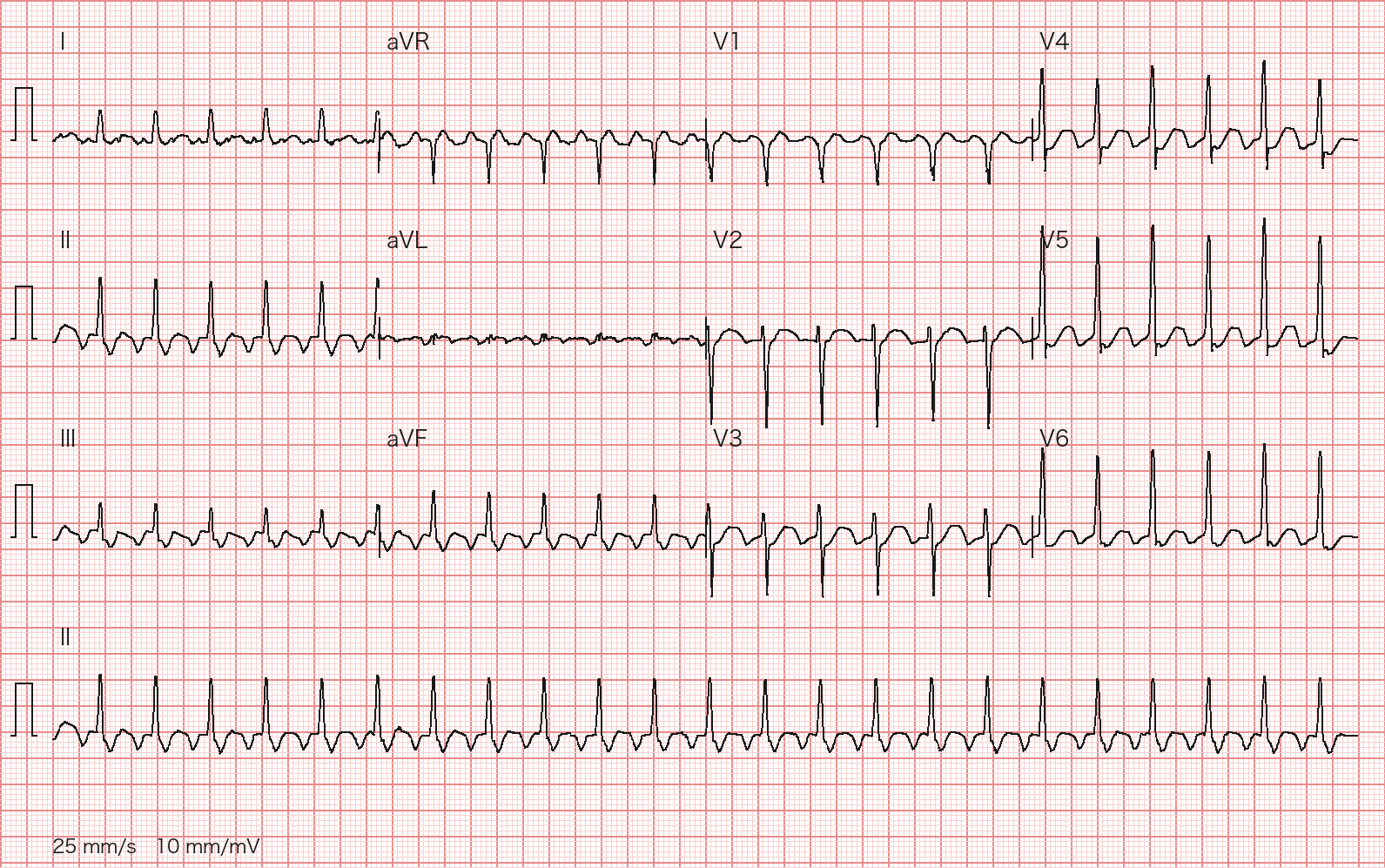
A 75-year-old woman has had palpitations for about 2 hours. BP 118/76 mm Hg, SpO2 97%. Her 12-lead ECG is shown. What is the rhythm?
Wagner P, Strodthoff N et al., PTB-XL 1.0.3, PhysioNet, CC BY 4.0; redrawn by BenchClub from the raw waveform - AAV nodal reentrant tachycardia
- BOrthodromic AV reentrant tachycardia
- CSinus tachycardia
- DAtrial flutter with 2:1 AV conduction
- EFocal atrial tachycardia with 1:1 conduction
Show answer
Answer D
Source PTB-XL cardiologist report (ecg_id 3306: atrial flutter with 2:1 AV block and rapid ventricular response); 2015 ACC/AHA/HRS Guideline for the Management of Adult Patients with Supraventricular Tachycardia
- Q02Multiple choiceCardiologyAdvanced
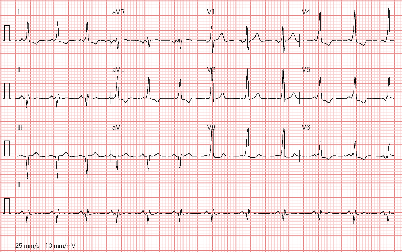
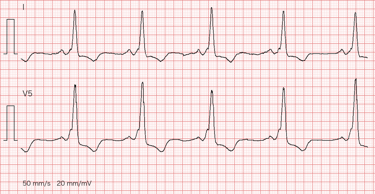
A 63-year-old man has a routine ECG before elective hernia repair. He has no chest pain or palpitations and his exercise tolerance is normal. His ECG is shown. Which interpretation is best supported?
Wagner P, Strodthoff N et al., PTB-XL 1.0.3, PhysioNet, CC BY 4.0; redrawn by BenchClub from the raw waveform Wagner P, Strodthoff N et al., PTB-XL 1.0.3, PhysioNet, CC BY 4.0; redrawn by BenchClub from the raw waveform; enlarged strip of leads I and V5 (50 mm/s, 20 mm/mV) - AOld inferior myocardial infarction
- BVentricular pre-excitation (Wolff-Parkinson-White pattern)
- CRight bundle branch block with left posterior fascicular block
- DRight ventricular hypertrophy
- EHypertrophic cardiomyopathy with septal hypertrophy
Show answer
Answer B
Source PTB-XL cardiologist report (ecg_id 12559: PR rather short but >0.12 s, delta waves evident, probable WPW); 2015 ACC/AHA/HRS Guideline for the Management of Adult Patients with Supraventricular Tachycardia (section 6, accessory pathways)
- Q03Multiple choiceCardiologyAdvanced
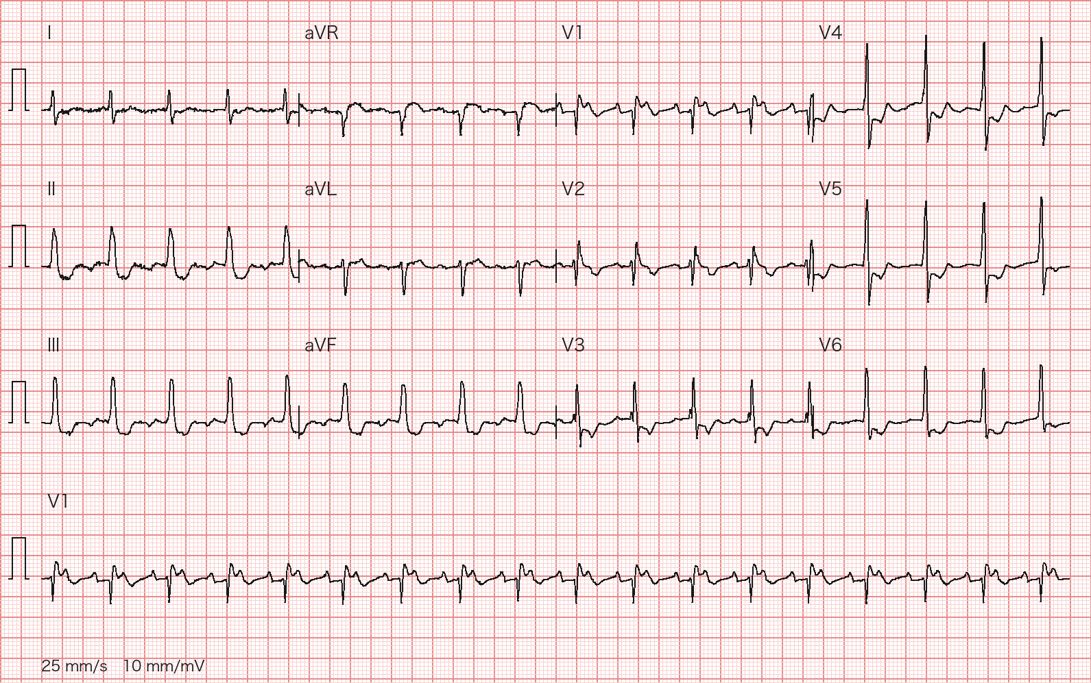
A 92-year-old woman is brought in with fatigue and dizziness. BP 104/62 mm Hg. Her ECG is shown; the rhythm strip at the bottom is lead V1. What is the rhythm?
Wagner P, Strodthoff N et al., PTB-XL 1.0.3, PhysioNet, CC BY 4.0; redrawn by BenchClub from the raw waveform - ASinus tachycardia with first-degree AV block
- BAtrial fibrillation with rapid ventricular response
- CAtrial tachycardia with 2:1 AV block
- DJunctional tachycardia
- ESinus rhythm with 2:1 second-degree AV block
Show answer
Answer C
Source PTB-XL cardiologist report (ecg_id 9136: atrial tachycardia with 2:1 AV block, incomplete RBBB, ST-T changes from ischaemia and digitalis effect); Goldberger's Clinical Electrocardiography, 10th ed.
- Q04Short answerCardiologyAdvanced
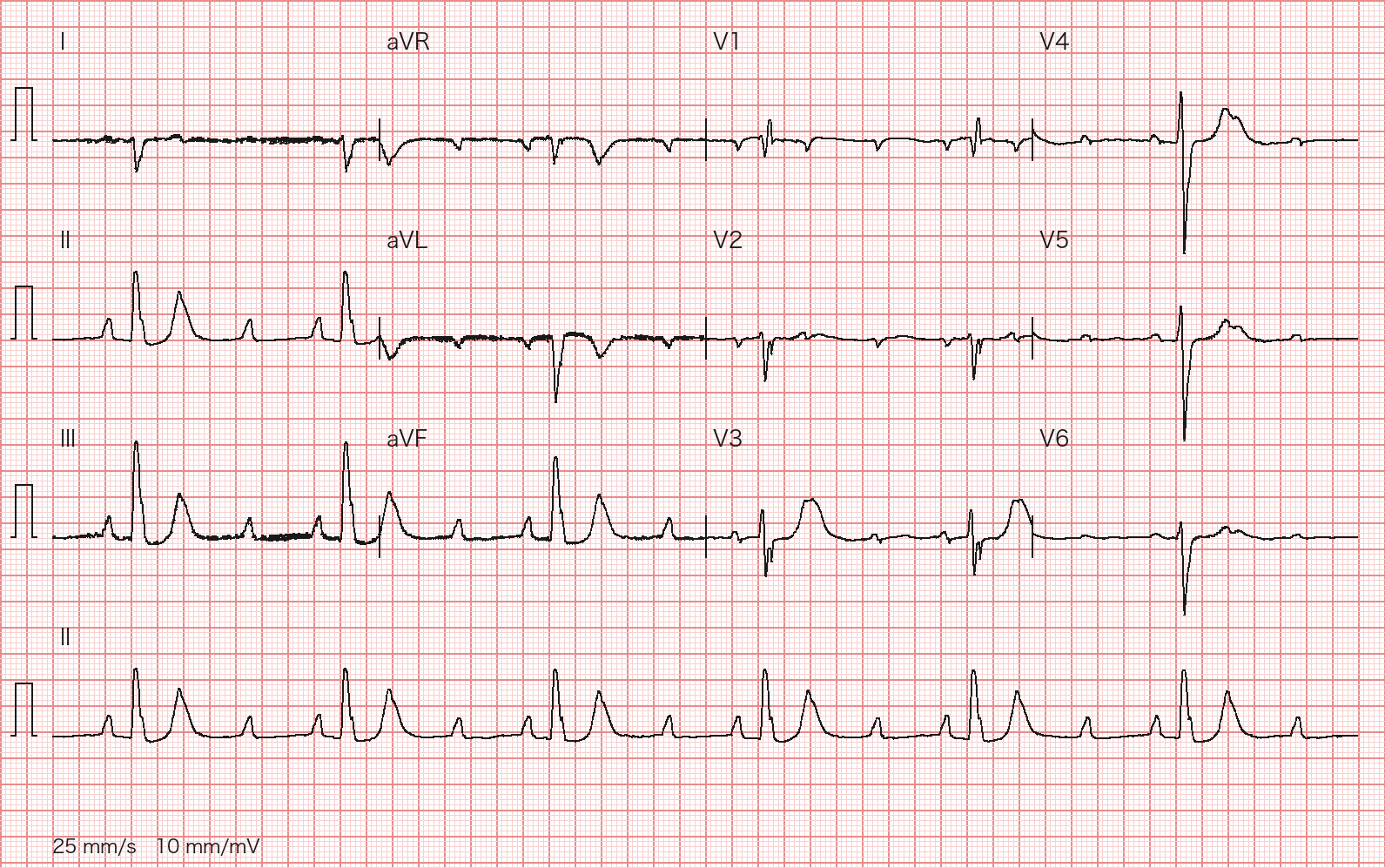
A 58-year-old man has had two episodes of near-syncope while climbing stairs in the past week. He takes no medications. BP 128/70 mm Hg; he is alert. Potassium and TSH are normal, and troponin is negative. His ECG is shown. Interpret the ECG systematically (rhythm and AV conduction, axis), classify the AV block, and give the definitive management.
Wagner P, Strodthoff N et al., PTB-XL 1.0.3, PhysioNet, CC BY 4.0; redrawn by BenchClub from the raw waveform Rubric
- +3Identifies 3:1 AV conduction (two consecutive nonconducted P waves) and classifies it as high-grade (advanced) second-degree AV block
- +1Gives an atrial rate of about 110/min and a ventricular rate of about 37/min
- +1Identifies right axis deviation
- +1Names left posterior fascicular block as a likely cause (accept LPFB versus RVH or right heart strain)
- +1Notes that the block is likely infranodal or that atropine is unlikely to help (or that the level cannot be fixed from the surface ECG)
- +2Recommends permanent pacemaker implantation
- +1Adds monitoring with transcutaneous pacing pads (or temporary pacing) while awaiting the implant
- -3Calls it Mobitz I (Wenckebach) or a benign vagal block and recommends observation only
Show answer
Reference answer Sinus tachycardia at about 110/min with 3:1 AV conduction: two consecutive P waves are not conducted for every one that is (one of them sits on or in the T wave, so the rhythm can be mistaken for 2:1), giving a ventricular rate of about 37/min. Two or more consecutive nonconducted P waves define high-grade (advanced) second-degree AV block; it is not Wenckebach. The axis is rightward (QRS negative in I, positive in II, III and aVF), read by the cardiologist as left posterior fascicular block. High-grade AV block together with fascicular disease favors infranodal (His-Purkinje) block, which is unlikely to respond to atropine and can progress abruptly to complete heart block. High-grade AV block without a reversible cause is a Class I indication for a permanent pacemaker, with or without symptoms and whatever the level; until implantation keep him monitored with transcutaneous pacing pads, with temporary pacing or a chronotropic infusion if he becomes unstable.
Source PTB-XL cardiologist report (ecg_id 8048: 3:1 AV block, rightward axis, LPFB, pacemaker advised); 2018 ACC/AHA/HRS Guideline on the Evaluation and Management of Patients With Bradycardia and Cardiac Conduction Delay
- Q05Short answerCardiologyAdvanced
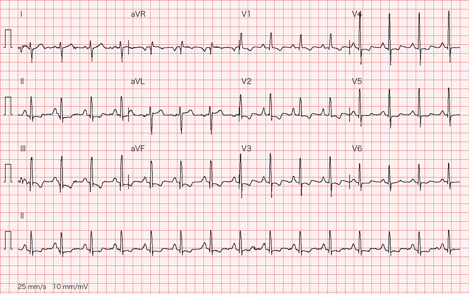
A 33-year-old man has 8 months of progressive exertional dyspnea and one episode of near-syncope while running for a bus. He does not smoke. SpO2 95% on room air. His ECG is shown. Describe the ECG findings, say what they point to, and outline the next diagnostic steps.
Wagner P, Strodthoff N et al., PTB-XL 1.0.3, PhysioNet, CC BY 4.0; redrawn by BenchClub from the raw waveform Rubric
- +2Identifies right atrial enlargement or abnormality (tall peaked P waves in the inferior leads)
- +2Identifies right axis deviation
- +2Identifies right ventricular hypertrophy (dominant R wave in V1, R/S > 1 in V1)
- +2Attributes the RAE, right axis and RVH pattern to RV pressure or volume overload, naming pulmonary hypertension (or atrial septal defect or pulmonary stenosis) as the leading concern
- +2Orders transthoracic echocardiography as the next test
- +1Includes right heart catheterization to confirm the diagnosis
- +1Includes a V/Q scan to look for chronic thromboembolic pulmonary hypertension
- -2Calls the ECG normal or a normal variant and stops the workup
Show answer
Reference answer Sinus rhythm. Tall peaked P waves in II, III and aVF (right atrial enlargement, 'P pulmonale'). Right axis deviation (reported as abnormal right axis). Dominant R wave in V1 (R/S > 1, early precordial transition) with persistent S waves in V5-V6 and repolarization changes: right ventricular hypertrophy. In a young man with exertional dyspnea and exertional near-syncope this points to RV pressure overload, most importantly pulmonary hypertension; an atrial septal defect or pulmonary stenosis can give a similar ECG. Next: transthoracic echocardiography (peak tricuspid regurgitation velocity, RV size and function, shunts or left heart disease), with BNP/NT-proBNP; if PH is likely, a V/Q scan to look for chronic thromboembolic disease and referral to a PH center for right heart catheterization to confirm and classify it (2022 ESC/ERS: mean pulmonary arterial pressure >20 mm Hg, pre-capillary if PAWP ≤15 mm Hg and PVR >2 Wood units).
Source PTB-XL cardiologist report (ecg_id 21050); 2022 ESC/ERS Guidelines for the Diagnosis and Treatment of Pulmonary Hypertension
- Q06Multiple choiceRadiologyAdvanced
An 87-year-old man in the ICU had an unsuccessful right subclavian central line attempt an hour ago. A portable radiograph is taken in bed (shown). What is the best interpretation and action?
Hellerhoff, Wikimedia Commons, CC BY-SA 4.0; re-encoded by BenchClub - ASmall right iatrogenic pneumothorax; small-bore chest tube on the right
- BSupine pneumothorax with a deep sulcus sign; small-bore chest tube
- CSkin folds plus a small true right apical pneumothorax; observe and repeat film
- DBullous emphysema, not pneumothorax; no drain, confirm with CT
- ESkin folds, not pneumothorax; no drain, confirm with lung ultrasound if doubt remains
Show answer
Answer E
Source Author's case description (Hellerhoff, Wikimedia Commons: skin folds on a bedside film, told apart from pneumothorax by their number); Felson's Principles of Chest Roentgenology, 5th ed.; BTS Guideline for Pleural Disease 2023
- Q07Multiple choiceRadiologyAdvanced
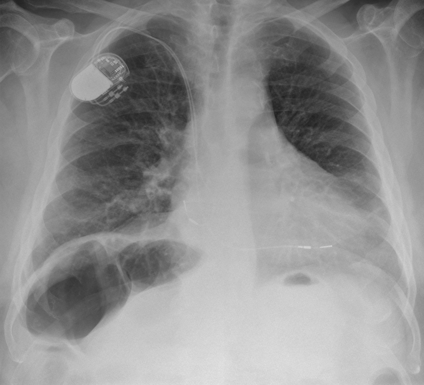
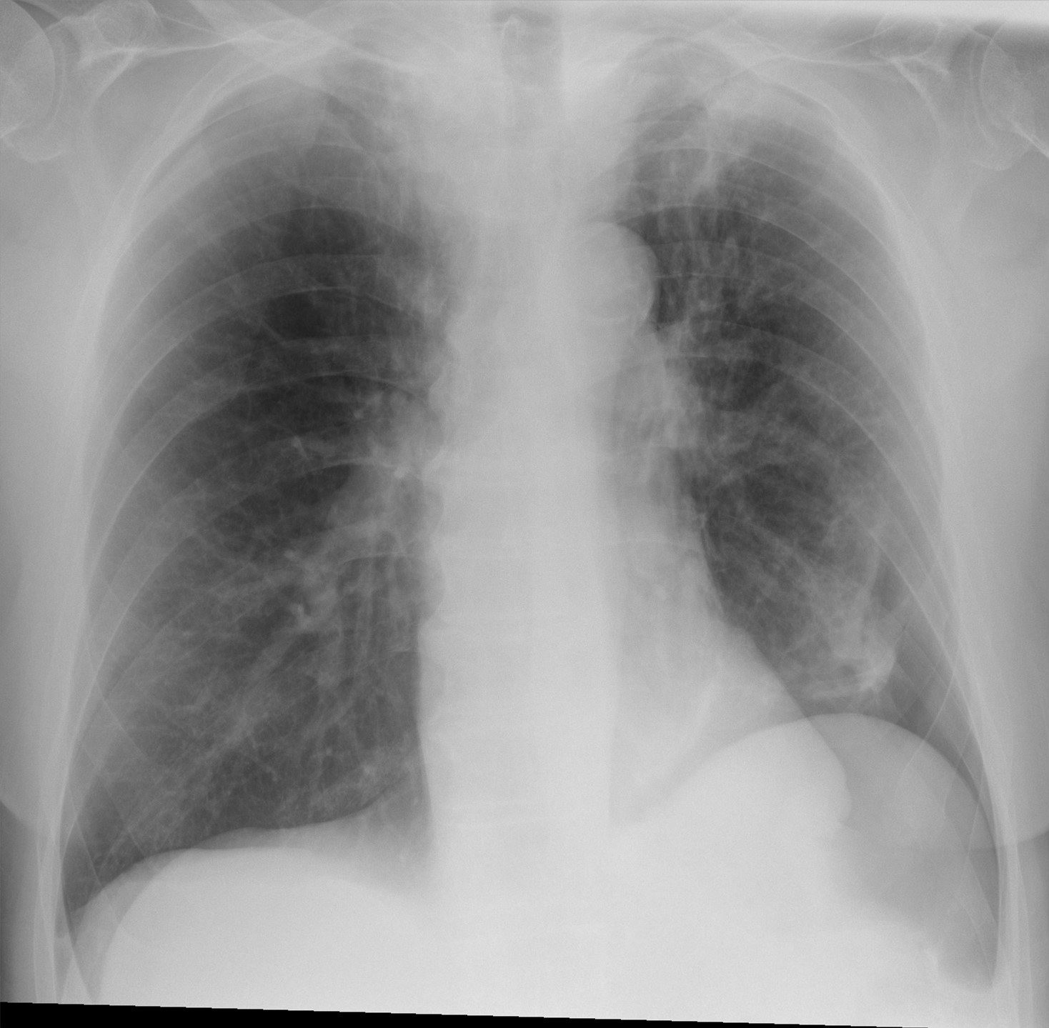
A 73-year-old man had a colonoscopy with removal of two polyps yesterday. Today he reports bloating and mild upper abdominal discomfort. An upright chest radiograph is shown, and the on-call surgeon asks for your reading. What is the best interpretation and next step?
Hellerhoff, Wikimedia Commons, CC BY-SA 4.0; mouse-cursor overlay removed and re-encoded by BenchClub - AFree intraperitoneal air from a colonic perforation; emergency laparotomy
- BColon interposed beneath the right hemidiaphragm (Chilaiditi sign); no emergency surgery
- CSubphrenic abscess; CT and percutaneous drainage
- DSmall free air from a contained microperforation; nonoperative care with serial examinations
- ERight basal pneumothorax; small-bore chest tube
Show answer
Answer B
Source Author's case description (Hellerhoff, Wikimedia Commons: Chilaiditi sign with distended hepatic flexure between diaphragm and liver); Felson's Principles of Chest Roentgenology, 5th ed.
- Q08Multiple choiceRadiologyAdvanced
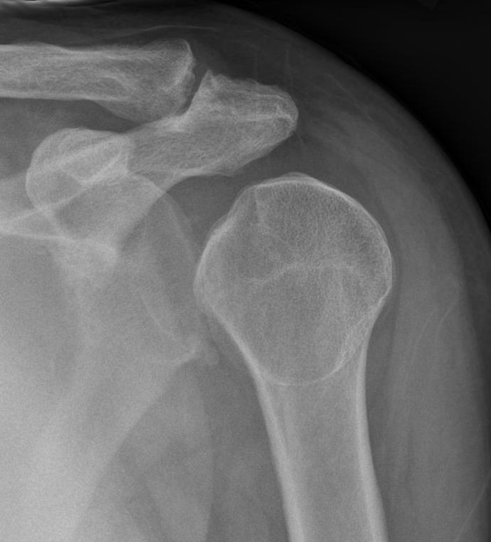
A 40-year-old man fell from his bicycle and has shoulder pain. Neurovascular examination is normal. A single AP radiograph of the shoulder is shown. What is the best interpretation?
Hellerhoff, Wikimedia Commons, CC BY-SA 3.0; pre-reduction panel cropped and re-encoded by BenchClub - ANo fracture or dislocation
- BAnterior (subcoracoid) glenohumeral dislocation
- CPosterior glenohumeral dislocation
- DImpacted surgical neck fracture of the humerus
- EInferior dislocation (luxatio erecta)
Show answer
Answer C
Source Author's case description (Hellerhoff, Wikimedia Commons: posterior shoulder dislocation with the lightbulb sign); Rockwood and Green's Fractures in Adults, 10th ed.
- Q09Short answerRadiologyAdvanced
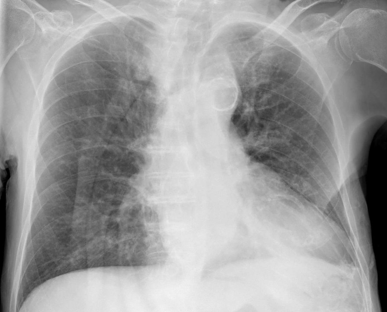
An 82-year-old man admitted for investigation of a pleural effusion becomes more short of breath on the ward: RR 26, SpO2 has fallen from 96% to 89% on 2 L of oxygen, BP 132/78 mm Hg. A portable supine AP radiograph is taken in bed (shown). Describe the key finding(s), give the diagnosis, and state your management.
Hellerhoff, Wikimedia Commons, CC BY-SA 4.0; mouse-cursor overlay removed and re-encoded by BenchClub Rubric
- +2Identifies a pneumothorax on the left side
- +2Names the deep sulcus sign (deep, lucent left costophrenic angle)
- +1Explains that on a supine film pleural air collects anteriorly and inferiorly, so an apical edge may be absent and the size is underestimated
- +1Notes the visceral pleural line visible at the left base
- +1Asks about or names a recent pleural procedure (thoracentesis) as the likely cause
- +2Recommends a small-bore chest drain (14 F or smaller) on the left with supplemental oxygen
- -3Misses the pneumothorax (says there is none or calls the lucency normal anatomy)
- -2Places the drain or locates the pneumothorax on the right
Show answer
Reference answer Left pneumothorax on a supine film. The left costophrenic sulcus is abnormally deep and lucent (deep sulcus sign) because pleural air rises to the anterior and inferior pleural space when the patient lies flat; there is no classic apical edge, but the visceral pleural line can be seen at the left base. Supine films underestimate pneumothorax size. In an inpatient with a pleural effusion the commonest cause is iatrogenic (recent thoracentesis), so ask about procedures. Significant hypoxemia (with or without underlying lung disease) is a high-risk feature in the BTS 2023 algorithm, so he needs supplemental oxygen and a small-bore (≤14 F) chest drain on the left rather than observation or simple aspiration; tension physiology is absent, so emergency needle decompression is not needed. If the lung fails to re-expand, consider trapped lung.
Source Author's case description (Hellerhoff, Wikimedia Commons: deep sulcus sign on a supine film, left pneumothorax after diagnostic pleural puncture, lung edge visible at the base); BTS Guideline for Pleural Disease 2023
- Q10Short answerRadiologyAdvanced
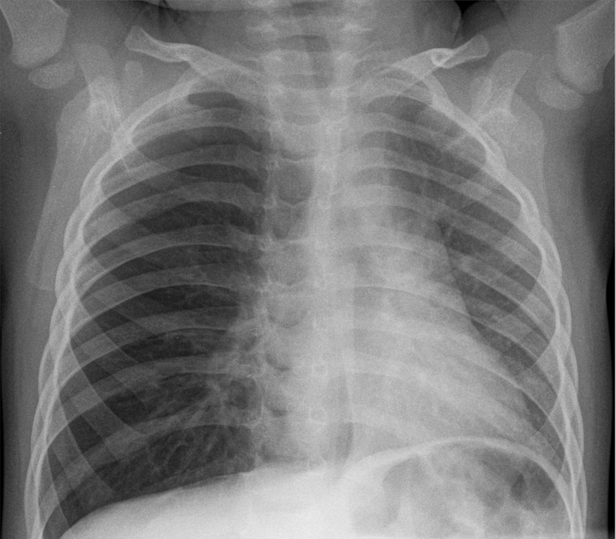
A 2-year-old boy has had 2 days of cough and intermittent wheeze. He has no fever. His parents do not recall a choking episode. Albuterol at an urgent care clinic did not help. RR 36, SpO2 94%. The frontal chest radiograph is shown. Describe the findings, give the most likely diagnosis and its mechanism, and state the next step.
Hellerhoff, Wikimedia Commons, CC BY-SA 4.0; bottom edge cropped and re-encoded by BenchClub Rubric
- +3Identifies hyperlucency and hyperexpansion of the right lung
- +2Identifies mediastinal shift to the left
- +2Names foreign-body aspiration into the right bronchial tree
- +1Explains the check-valve (ball-valve) mechanism of air trapping
- +2Recommends bronchoscopy to find and remove the foreign body (rigid bronchoscopy under general anesthesia is standard)
- -2Places the hyperlucent, air-trapped lung or the foreign body on the left
- -2Manages it as asthma, bronchiolitis or pneumonia without airway evaluation
Show answer
Reference answer The right lung is hyperlucent and overexpanded (more rib spaces, flattened right hemidiaphragm) with mediastinal shift to the left, away from the lucent side. In a toddler with new cough and wheeze that does not respond to bronchodilators this is unilateral air trapping from a check-valve (ball-valve) obstruction: an aspirated foreign body (often a nut or food) in the right bronchus lets air in on inspiration but not out on expiration. The right lung is the abnormal one: it is overexpanded and oligemic, while the left lung is aerated and not collapsed, which separates right-sided air trapping from left collapse with compensatory hyperinflation. A negative choking history does not exclude it. Expiratory or bilateral decubitus films can show the trapping (the dependent lucent lung does not deflate) but must not delay treatment. Next step: rigid bronchoscopy under general anesthesia to confirm and remove the foreign body.
Source Author's case description (Hellerhoff, Wikimedia Commons: foreign-body aspiration with check-valve mechanism, overinflation of the right lung and mediastinal shift to the left); Nelson Textbook of Pediatrics, 22nd ed. (foreign bodies in the airway)
- Q11Short answerRadiologyAdvanced
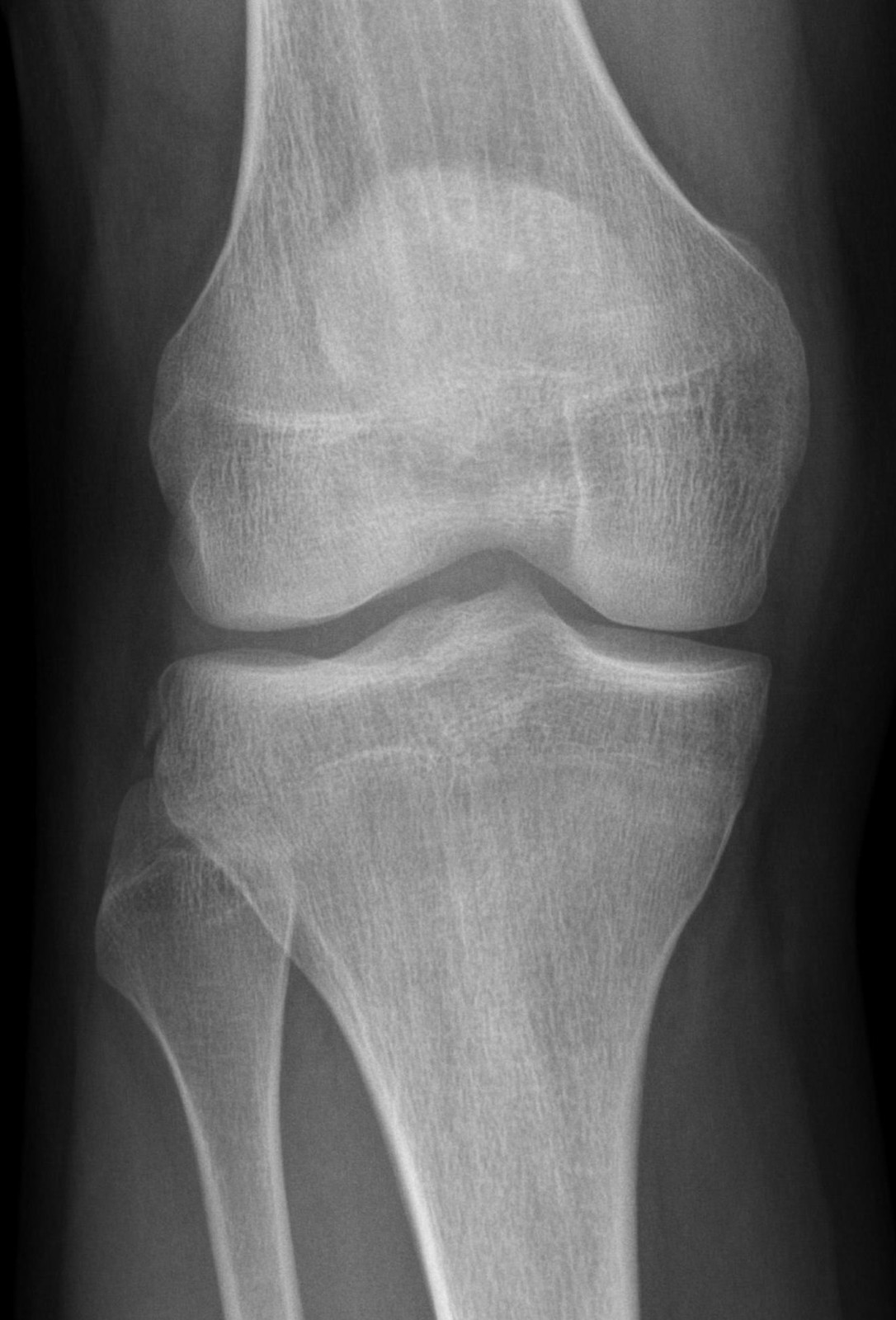
An 18-year-old man injured his knee yesterday. He can bear weight with pain. The AP radiograph is shown. Describe the radiograph findings, what they imply, and the next steps.
Hellerhoff, Wikimedia Commons, CC BY-SA 4.0; downscaled and re-encoded by BenchClub Rubric
- +2Identifies the small avulsion fragment at the lateral margin of the lateral tibial plateau
- +2Names it a Segond fracture (lateral capsular sign; anterolateral ligament or lateral capsular avulsion accepted)
- +2States its strong association with anterior cruciate ligament tear
- +1Mentions associated meniscal or other ligament injury
- +2Recommends MRI of the knee
- +1Recommends orthopedic or sports-medicine referral
- -3Reads the radiograph as normal and discharges with symptomatic care only
- -2Calls it a reverse Segond (medial) fracture or links it to a posterior cruciate ligament tear
Show answer
Reference answer There is a small, elliptical avulsion fragment at the lateral margin of the lateral tibial plateau, just below the joint line: a Segond fracture (lateral capsular sign), an avulsion at the attachment of the lateral capsule/anterolateral ligament from internal rotation and varus stress. It is strongly associated with anterior cruciate ligament rupture (most reported series 75-100%) and often with meniscal injury. Next: examine for ACL laxity (Lachman), protect the knee (brace, crutches), obtain an MRI of the knee to assess the ACL, menisci and other ligaments, and refer to orthopedics/sports medicine.
Source Author's case description (Hellerhoff, Wikimedia Commons: Segond fracture with rupture of the anterior cruciate ligament); ACR Appropriateness Criteria Acute Trauma to the Knee (2019)
- Q12Multiple choiceDermatologyAdvanced
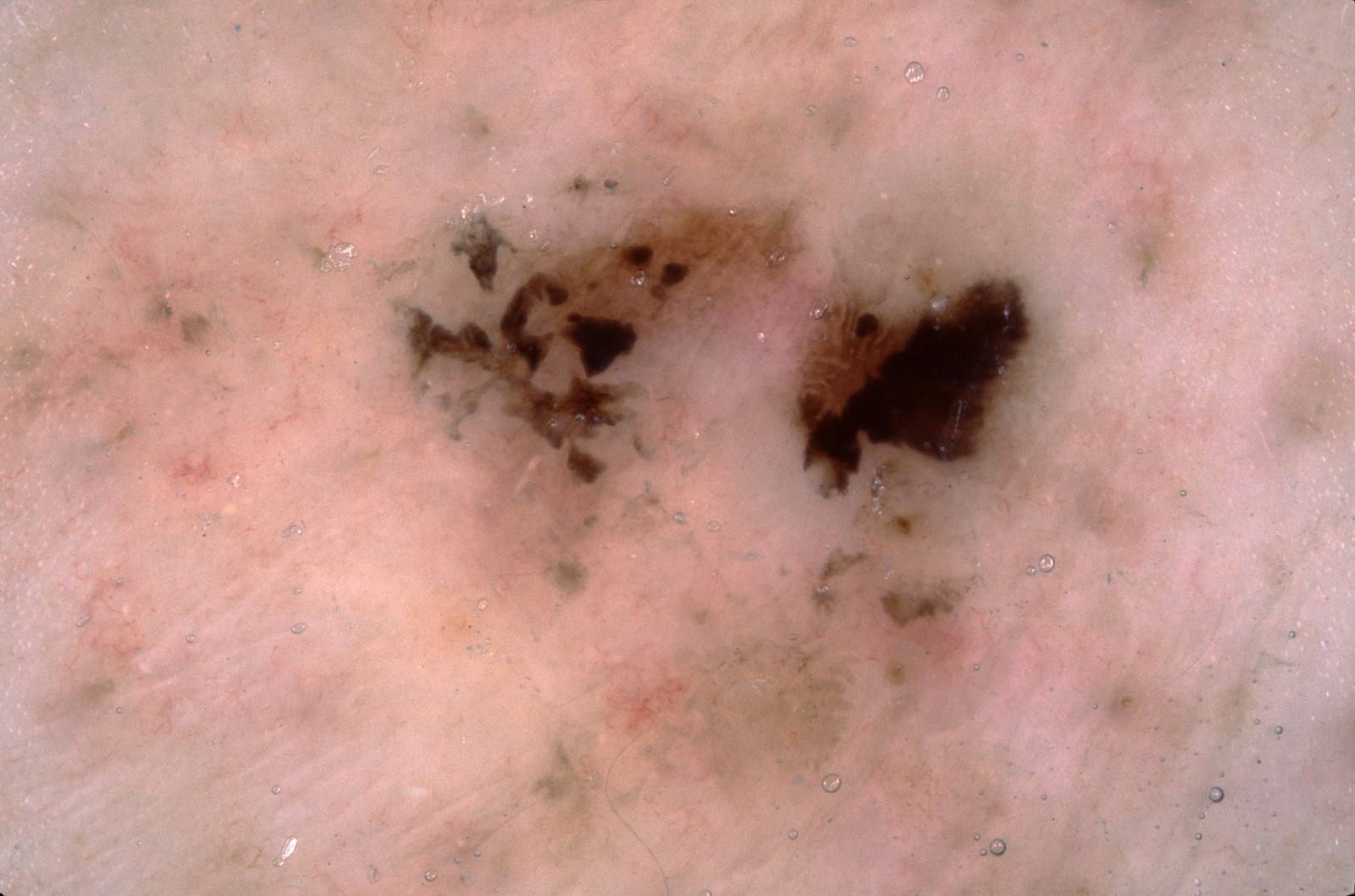
An 85-year-old man has a flat, multicolored lesion on his chest. The dermoscopic image is shown; the lesion was biopsied. What is the most likely histopathologic diagnosis?
Anonymous contributor, ISIC Archive ISIC_0012070, CC0 - ASuperficial spreading melanoma
- BSeborrheic keratosis
- CPigmented Bowen disease (squamous cell carcinoma in situ)
- DPigmented superficial basal cell carcinoma
- ELichen planus-like keratosis
Show answer
Answer D
Source ISIC Archive histopathology label (Basal cell carcinoma, superficial); Kittler H et al., Standardization of terminology in dermoscopy/dermatoscopy, JAAD 2016; Lallas A et al., The dermatoscopic universe of basal cell carcinoma (Dermatol Pract Concept 2014)
- Q13Short answerDermatologyAdvanced
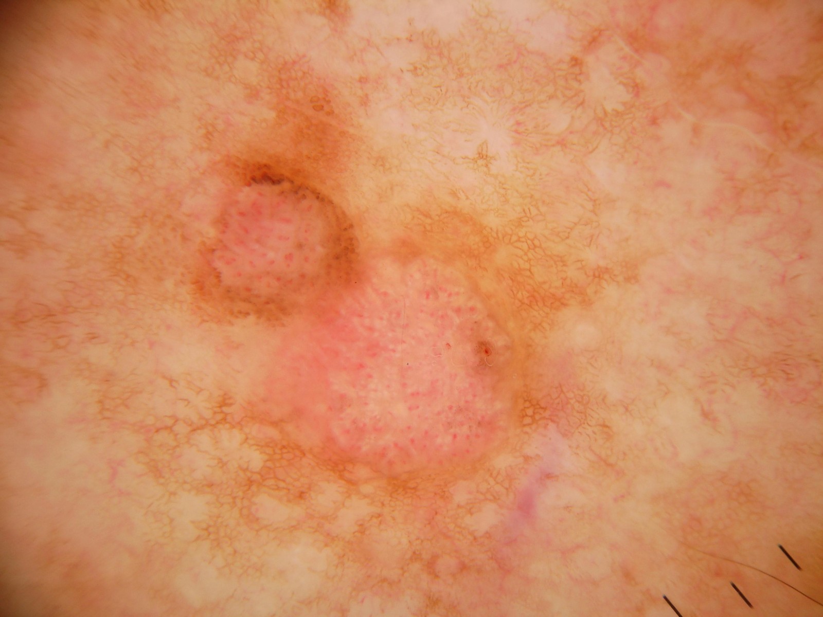
A 70-year-old man has two adjacent pink plaques on his shoulder; he is not sure how long they have been there. The dermoscopic image is shown. Give the most likely diagnosis, the dermoscopic clues that support it, the main differential, and how you would manage it.
Anonymous contributor, ISIC Archive ISIC_0022443, CC0 Rubric
- +3Diagnoses squamous cell carcinoma in situ (Bowen disease) as the most likely diagnosis
- +2Cites dotted or glomerular (coiled) vessels, diffuse or clustered, as the key dermoscopic clue
- +1Notes the peripheral brown dots or pigmentation at the lesion edge
- +1Names superficial basal cell carcinoma in the differential
- +1Confirms the diagnosis with a biopsy
- +2Offers appropriate treatments for SCC in situ (excision, curettage and electrodesiccation, cryotherapy, topical 5-fluorouracil or imiquimod, or photodynamic therapy)
- -3Gives melanoma or amelanotic melanoma as the most likely diagnosis
- -2Treats it as dermatitis or psoriasis with a topical corticosteroid, or recommends observation without biopsy
Show answer
Reference answer Squamous cell carcinoma in situ (Bowen disease), which histopathology confirmed. Clues: dotted and glomerular (coiled) vessels spread over the pink areas, with brown dots and pigmentation at the edge of the upper plaque, in two adjacent pink plaques on sun-damaged skin (background solar lentigines). Differential: superficial basal cell carcinoma (short fine telangiectasias, leaf-like areas, shiny white structures), psoriasis or eczema (regularly distributed dotted vessels), actinic keratosis and amelanotic melanoma. Confirm with a shave or punch biopsy. SCC in situ is low-risk: options are excision, curettage and electrodesiccation, cryotherapy, or topical 5-fluorouracil or imiquimod or photodynamic therapy for selected lesions; arrange skin surveillance.
Source ISIC Archive histopathology label (Squamous cell carcinoma in situ, Bowen disease); Zalaudek I et al., Dermoscopy of Bowen's disease (Br J Dermatol 2004); AAD Guidelines of care for the management of cutaneous squamous cell carcinoma (Alam M et al., JAAD 2018)
- Q14Short answerDermatologyAdvanced
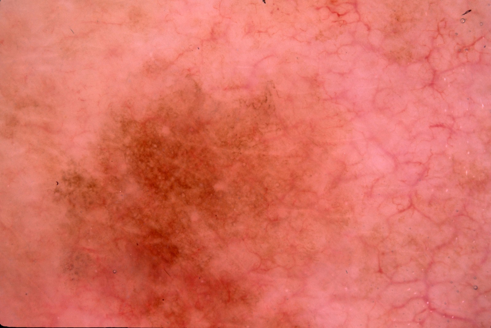
An 85-year-old woman has a flat lesion on her upper chest. The dermoscopic image is shown. Describe the dermoscopic findings, give the most likely diagnosis and the most important differential, and state what you would do.
Anonymous contributor, ISIC Archive ISIC_0012052, CC0 Rubric
- +2Describes gray or brown fine granularity (peppering)
- +1Describes the reddish background with branching vessels of sun-damaged skin
- +2Names lichen planus-like keratosis (benign lichenoid keratosis) as the most likely diagnosis or as a close second to melanoma in situ
- +2Names melanoma in situ or lentigo maligna as the differential that cannot be excluded
- +2Recommends a biopsy (broad shave, saucerization or excision) for histopathology
- -3Reassures or discharges with no histology
- -2Recommends wide excision with melanoma margins or sentinel node biopsy before histology
Show answer
Reference answer Reddish background with prominent branching vessels of photodamaged skin, predominantly brown, partly gray fine granularity (peppering) and a few brown structureless and reticular areas, without a well-formed pigment network or melanoma-specific vessels: the pattern of a regressing lichenoid keratosis, i.e. lichen planus-like keratosis (benign lichenoid keratosis), which histopathology confirmed. The most important differential is melanoma in situ (lentigo maligna type on sun-damaged skin), because gray granularity and regression also occur in melanoma; pigmented actinic keratosis is another. A dermoscopic diagnosis of LPLK is not safe enough to reassure an 85-year-old with a solitary lesion of this kind: perform a biopsy (broad shave/saucerization or excision) for histopathology.
Source ISIC Archive histopathology label (Lichen planus-like keratosis); Bugatti L, Filosa G, Dermoscopy of lichen planus-like keratosis (J Eur Acad Dermatol Venereol 2007); Kittler H et al., JAAD 2016
- Q15Multiple choiceDermatologyAdvanced
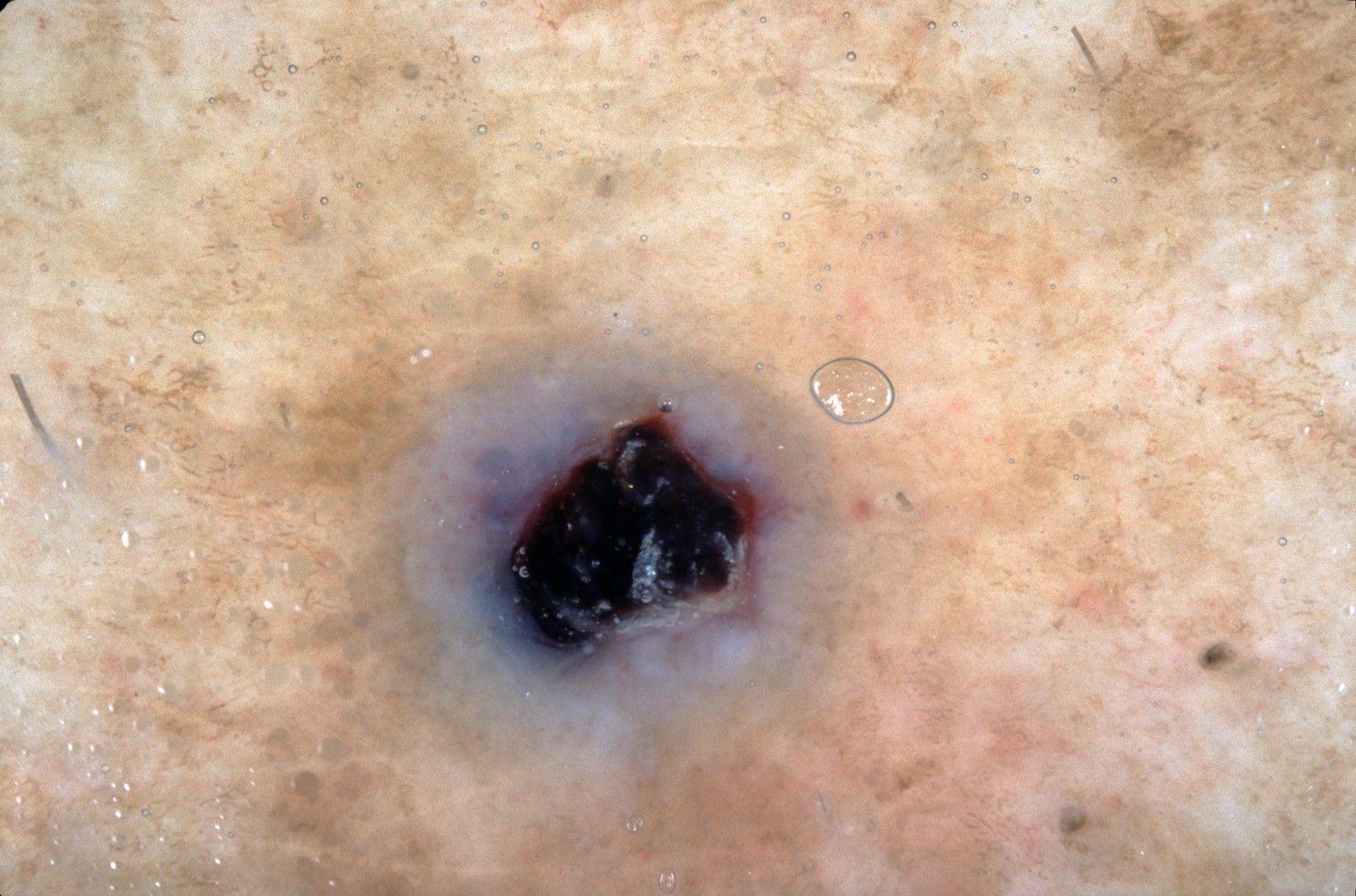
A 70-year-old woman has a pigmented lesion on her leg that bled after a minor knock. The dermoscopic image is shown; the lesion was excised. What is the most likely histopathologic diagnosis?
Anonymous contributor, ISIC Archive ISIC_0015306, CC0 - AAngiokeratoma
- BDermatofibroma
- CBlue nevus
- DPigmented basal cell carcinoma
- ESeborrheic keratosis
Show answer
Answer A
Source ISIC Archive histopathology label (Angiokeratoma); Zaballos P et al., Dermoscopy of solitary angiokeratomas (Arch Dermatol 2007); Kittler H et al., JAAD 2016
- Q16Short answerDermatologyAdvanced
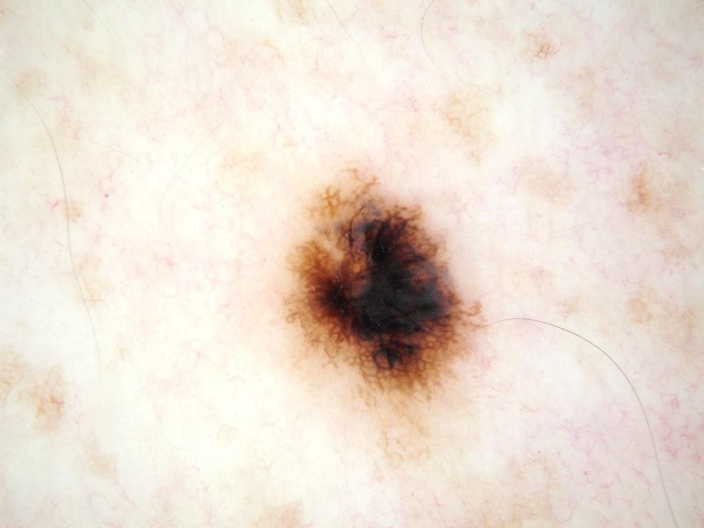
A 45-year-old man noticed a new spot about 5 mm across on his chest 4 months ago. The dermoscopic image is shown. Describe the dermoscopic features, give your differential diagnosis, and state the management.
Anonymous contributor, ISIC Archive ISIC_0010814, CC0 Rubric
- +1Describes the central or eccentric black blotch
- +1Describes the blue-gray or whitish veil-like area
- +1Describes the irregular or thickened peripheral network, or the asymmetry
- +2Names melanoma as the main diagnosis to exclude and ties it to a visible feature
- +1Names a benign alternative (Reed/Spitz nevus or atypical nevus)
- +2Recommends complete removal for histopathology with 1-3 mm clinical margins (excisional biopsy or deep saucerization), not a superficial shave
- -3Recommends reassurance or digital dermoscopic monitoring instead of removal
- -2Recommends wide local excision with 1-2 cm margins or sentinel node biopsy before histology
Show answer
Reference answer An asymmetric lesion with an eccentric black blotch, a blue-gray to whitish veil-like area and an irregular, thickened peripheral network. These are melanoma-associated features, so melanoma is the diagnosis to exclude. A pigmented spindle cell (Reed) nevus, the final histopathologic diagnosis here, or an atypical nevus can look like this, but in an adult with a new lesion dermoscopy cannot separate them from melanoma. Management: complete removal for histopathology with 1-3 mm clinical margins (excisional biopsy, or a deep saucerization that removes the whole lesion); no monitoring, and no wide excision or sentinel node biopsy before the diagnosis is known.
Source ISIC Archive histopathology label (Nevus, Reed); Lallas A et al., Update on dermoscopy of Spitz/Reed naevi and management guidelines by the International Dermoscopy Society (Br J Dermatol 2017); AAD Guidelines of care for the management of primary cutaneous melanoma (Swetter SM et al., JAAD 2019)